Chlorine »
PDB 1nc7-1nvf »
1nvd »
Chlorine in PDB 1nvd: Crystal Structure of 3-Dehydroquinate Synthase (Dhqs) in Complex with ZN2+ and Carbaphosphonate
Enzymatic activity of Crystal Structure of 3-Dehydroquinate Synthase (Dhqs) in Complex with ZN2+ and Carbaphosphonate
All present enzymatic activity of Crystal Structure of 3-Dehydroquinate Synthase (Dhqs) in Complex with ZN2+ and Carbaphosphonate:
4.2.3.4;
4.2.3.4;
Protein crystallography data
The structure of Crystal Structure of 3-Dehydroquinate Synthase (Dhqs) in Complex with ZN2+ and Carbaphosphonate, PDB code: 1nvd
was solved by
C.E.Nichols,
J.Ren,
H.K.Lamb,
A.R.Hawkins,
D.K.Stammers,
with X-Ray Crystallography technique. A brief refinement statistics is given in the table below:
| Resolution Low / High (Å) | 30.00 / 2.51 |
| Space group | P 21 21 21 |
| Cell size a, b, c (Å), α, β, γ (°) | 64.030, 70.030, 197.630, 90.00, 90.00, 90.00 |
| R / Rfree (%) | 19.6 / 26.7 |
Other elements in 1nvd:
The structure of Crystal Structure of 3-Dehydroquinate Synthase (Dhqs) in Complex with ZN2+ and Carbaphosphonate also contains other interesting chemical elements:
| Zinc | (Zn) | 2 atoms |
Chlorine Binding Sites:
The binding sites of Chlorine atom in the Crystal Structure of 3-Dehydroquinate Synthase (Dhqs) in Complex with ZN2+ and Carbaphosphonate
(pdb code 1nvd). This binding sites where shown within
5.0 Angstroms radius around Chlorine atom.
In total only one binding site of Chlorine was determined in the Crystal Structure of 3-Dehydroquinate Synthase (Dhqs) in Complex with ZN2+ and Carbaphosphonate, PDB code: 1nvd:
In total only one binding site of Chlorine was determined in the Crystal Structure of 3-Dehydroquinate Synthase (Dhqs) in Complex with ZN2+ and Carbaphosphonate, PDB code: 1nvd:
Chlorine binding site 1 out of 1 in 1nvd
Go back to
Chlorine binding site 1 out
of 1 in the Crystal Structure of 3-Dehydroquinate Synthase (Dhqs) in Complex with ZN2+ and Carbaphosphonate
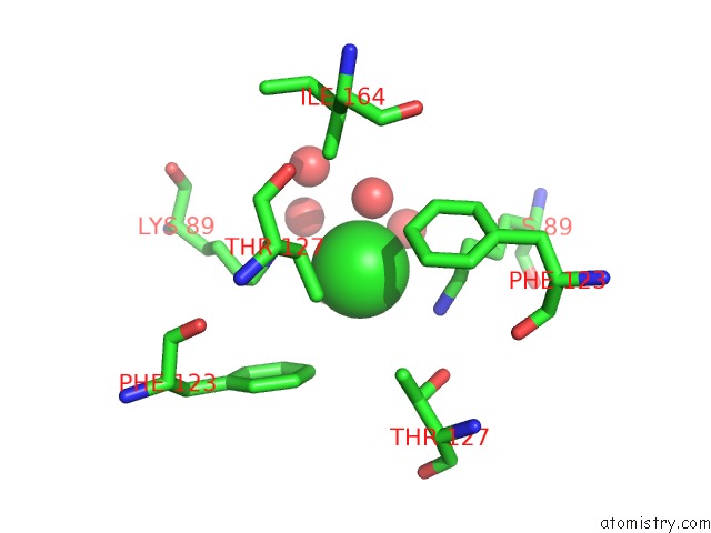
Mono view
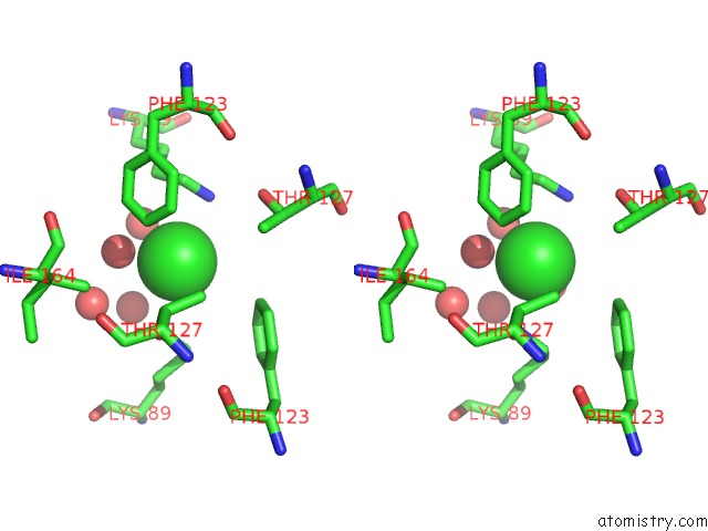
Stereo pair view
Mono view
Stereo pair view
A full contact list of Chlorine with other atoms in the Cl binding
site number 1 of Crystal Structure of 3-Dehydroquinate Synthase (Dhqs) in Complex with ZN2+ and Carbaphosphonate within 5.0Å range:
|
Reference:
C.E.Nichols,
J.Ren,
H.K.Lamb,
A.R.Hawkins,
D.K.Stammers.
Ligand-Induced Conformational Changes and A Mechanism For Domain Closure in Aspergillus Nidulans Dehydroquinate Synthase J.Mol.Biol. V. 327 129 2003.
ISSN: ISSN 0022-2836
PubMed: 12614613
DOI: 10.1016/S0022-2836(03)00086-X
Page generated: Sat Jul 20 00:39:57 2024
ISSN: ISSN 0022-2836
PubMed: 12614613
DOI: 10.1016/S0022-2836(03)00086-X
Last articles
Zn in 9J0NZn in 9J0O
Zn in 9J0P
Zn in 9FJX
Zn in 9EKB
Zn in 9C0F
Zn in 9CAH
Zn in 9CH0
Zn in 9CH3
Zn in 9CH1