Chlorine »
PDB 1nw5-1o54 »
1nzl »
Chlorine in PDB 1nzl: Crystal Structure of Src SH2 Domain Bound to Doubly Phosphorylated Peptide Pqpyepyipi
Enzymatic activity of Crystal Structure of Src SH2 Domain Bound to Doubly Phosphorylated Peptide Pqpyepyipi
All present enzymatic activity of Crystal Structure of Src SH2 Domain Bound to Doubly Phosphorylated Peptide Pqpyepyipi:
2.7.1.112;
2.7.1.112;
Protein crystallography data
The structure of Crystal Structure of Src SH2 Domain Bound to Doubly Phosphorylated Peptide Pqpyepyipi, PDB code: 1nzl
was solved by
O.Y.Lubman,
G.Waksman,
with X-Ray Crystallography technique. A brief refinement statistics is given in the table below:
| Resolution Low / High (Å) | 28.27 / 1.90 |
| Space group | P 21 21 2 |
| Cell size a, b, c (Å), α, β, γ (°) | 100.321, 68.455, 29.732, 90.00, 90.00, 90.00 |
| R / Rfree (%) | 22.3 / 26.5 |
Chlorine Binding Sites:
The binding sites of Chlorine atom in the Crystal Structure of Src SH2 Domain Bound to Doubly Phosphorylated Peptide Pqpyepyipi
(pdb code 1nzl). This binding sites where shown within
5.0 Angstroms radius around Chlorine atom.
In total 2 binding sites of Chlorine where determined in the Crystal Structure of Src SH2 Domain Bound to Doubly Phosphorylated Peptide Pqpyepyipi, PDB code: 1nzl:
Jump to Chlorine binding site number: 1; 2;
In total 2 binding sites of Chlorine where determined in the Crystal Structure of Src SH2 Domain Bound to Doubly Phosphorylated Peptide Pqpyepyipi, PDB code: 1nzl:
Jump to Chlorine binding site number: 1; 2;
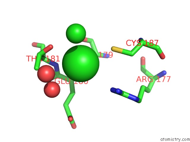
Chlorine binding site 1 out of 2 in 1nzl
Go back to
Chlorine binding site 1 out
of 2 in the Crystal Structure of Src SH2 Domain Bound to Doubly Phosphorylated Peptide Pqpyepyipi
Mono view
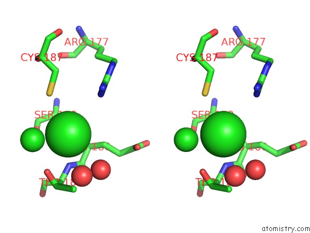
Stereo pair view
Mono view
Stereo pair view
A full contact list of Chlorine with other atoms in the Cl binding
site number 1 of Crystal Structure of Src SH2 Domain Bound to Doubly Phosphorylated Peptide Pqpyepyipi within 5.0Å range:
|
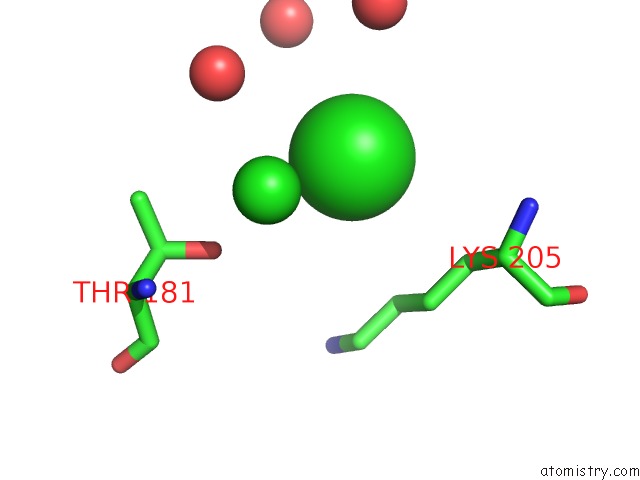
Chlorine binding site 2 out of 2 in 1nzl
Go back to
Chlorine binding site 2 out
of 2 in the Crystal Structure of Src SH2 Domain Bound to Doubly Phosphorylated Peptide Pqpyepyipi
Mono view
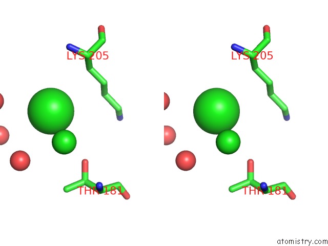
Stereo pair view
Mono view
Stereo pair view
A full contact list of Chlorine with other atoms in the Cl binding
site number 2 of Crystal Structure of Src SH2 Domain Bound to Doubly Phosphorylated Peptide Pqpyepyipi within 5.0Å range:
|
Reference:
O.Y.Lubman,
G.Waksman.
Structural and Thermodynamic Basis For the Interaction of the Src SH2 Domain with the Activated Form of the Pdgf Beta-Receptor J.Mol.Biol. V. 328 655 2003.
ISSN: ISSN 0022-2836
PubMed: 12706723
DOI: 10.1016/S0022-2836(03)00344-9
Page generated: Sat Jul 20 00:42:41 2024
ISSN: ISSN 0022-2836
PubMed: 12706723
DOI: 10.1016/S0022-2836(03)00344-9
Last articles
Zn in 9J0NZn in 9J0O
Zn in 9J0P
Zn in 9FJX
Zn in 9EKB
Zn in 9C0F
Zn in 9CAH
Zn in 9CH0
Zn in 9CH3
Zn in 9CH1