Chlorine »
PDB 1o5e-1omy »
1o6b »
Chlorine in PDB 1o6b: Crystal Structure of Phosphopantetheine Adenylyltransferase with Adp
Enzymatic activity of Crystal Structure of Phosphopantetheine Adenylyltransferase with Adp
All present enzymatic activity of Crystal Structure of Phosphopantetheine Adenylyltransferase with Adp:
2.7.7.3;
2.7.7.3;
Protein crystallography data
The structure of Crystal Structure of Phosphopantetheine Adenylyltransferase with Adp, PDB code: 1o6b
was solved by
Structural Genomix,
with X-Ray Crystallography technique. A brief refinement statistics is given in the table below:
| Resolution Low / High (Å) | 32.01 / 2.20 |
| Space group | P 41 3 2 |
| Cell size a, b, c (Å), α, β, γ (°) | 115.420, 115.420, 115.420, 90.00, 90.00, 90.00 |
| R / Rfree (%) | 22.9 / 27.5 |
Other elements in 1o6b:
The structure of Crystal Structure of Phosphopantetheine Adenylyltransferase with Adp also contains other interesting chemical elements:
| Magnesium | (Mg) | 1 atom |
Chlorine Binding Sites:
The binding sites of Chlorine atom in the Crystal Structure of Phosphopantetheine Adenylyltransferase with Adp
(pdb code 1o6b). This binding sites where shown within
5.0 Angstroms radius around Chlorine atom.
In total only one binding site of Chlorine was determined in the Crystal Structure of Phosphopantetheine Adenylyltransferase with Adp, PDB code: 1o6b:
In total only one binding site of Chlorine was determined in the Crystal Structure of Phosphopantetheine Adenylyltransferase with Adp, PDB code: 1o6b:
Chlorine binding site 1 out of 1 in 1o6b
Go back to
Chlorine binding site 1 out
of 1 in the Crystal Structure of Phosphopantetheine Adenylyltransferase with Adp

Mono view
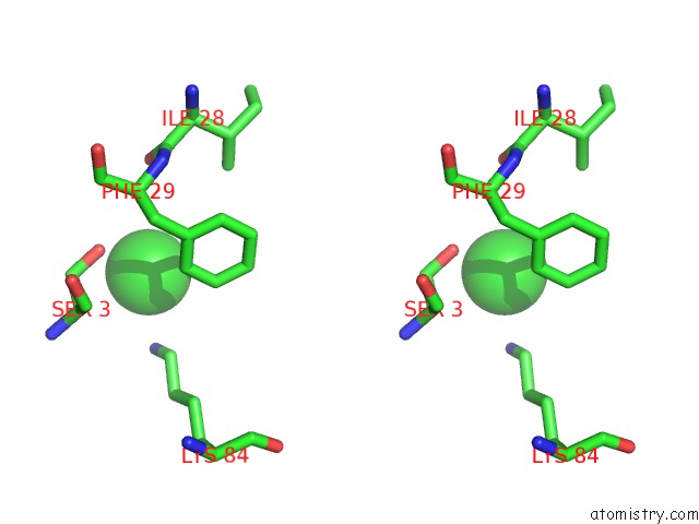
Stereo pair view
Mono view
Stereo pair view
A full contact list of Chlorine with other atoms in the Cl binding
site number 1 of Crystal Structure of Phosphopantetheine Adenylyltransferase with Adp within 5.0Å range:
|
Reference:
J.Badger,
J.M.Sauder,
J.M.Adams,
S.Antonysamy,
K.Bain,
M.G.Bergseid,
S.G.Buchanan,
M.D.Buchanan,
Y.Batiyenko,
J.A.Christopher,
S.Emtage,
A.Eroshkina,
I.Feil,
E.B.Furlong,
K.S.Gajiwala,
X.Gao,
D.He,
J.Hendle,
A.Huber,
K.Hoda,
P.Kearins,
C.Kissinger,
B.Laubert,
H.A.Lewis,
J.Lin,
K.Loomis,
D.Lorimer,
G.Louie,
M.Maletic,
C.D.Marsh,
I.Miller,
J.Molinari,
H.J.Muller-Dieckmann,
J.M.Newman,
B.W.Noland,
B.Pagarigan,
F.Park,
T.S.Peat,
K.W.Post,
S.Radojicic,
A.Ramos,
R.Romero,
M.E.Rutter,
W.E.Sanderson,
K.D.Schwinn,
J.Tresser,
J.Winhoven,
T.A.Wright,
L.Wu,
J.Xu,
T.J.Harris.
Structural Analysis of A Set of Proteins Resulting From A Bacterial Genomics Project Proteins V. 60 787 2005.
ISSN: ISSN 0887-3585
PubMed: 16021622
DOI: 10.1002/PROT.20541
Page generated: Sat Jul 20 00:49:11 2024
ISSN: ISSN 0887-3585
PubMed: 16021622
DOI: 10.1002/PROT.20541
Last articles
Zn in 9J0NZn in 9J0O
Zn in 9J0P
Zn in 9FJX
Zn in 9EKB
Zn in 9C0F
Zn in 9CAH
Zn in 9CH0
Zn in 9CH3
Zn in 9CH1