Chlorine »
PDB 1o5e-1omy »
1ogd »
Chlorine in PDB 1ogd: The Structure of Bacillus Subtilis Rbsd Complexed with D-Ribose
Protein crystallography data
The structure of The Structure of Bacillus Subtilis Rbsd Complexed with D-Ribose, PDB code: 1ogd
was solved by
M.-S.Kim,
B.-H.Oh,
with X-Ray Crystallography technique. A brief refinement statistics is given in the table below:
| Resolution Low / High (Å) | 30.00 / 1.95 |
| Space group | C 1 2 1 |
| Cell size a, b, c (Å), α, β, γ (°) | 123.515, 108.656, 83.308, 90.00, 128.68, 90.00 |
| R / Rfree (%) | 19.8 / 21.1 |
Chlorine Binding Sites:
The binding sites of Chlorine atom in the The Structure of Bacillus Subtilis Rbsd Complexed with D-Ribose
(pdb code 1ogd). This binding sites where shown within
5.0 Angstroms radius around Chlorine atom.
In total 2 binding sites of Chlorine where determined in the The Structure of Bacillus Subtilis Rbsd Complexed with D-Ribose, PDB code: 1ogd:
Jump to Chlorine binding site number: 1; 2;
In total 2 binding sites of Chlorine where determined in the The Structure of Bacillus Subtilis Rbsd Complexed with D-Ribose, PDB code: 1ogd:
Jump to Chlorine binding site number: 1; 2;
Chlorine binding site 1 out of 2 in 1ogd
Go back to
Chlorine binding site 1 out
of 2 in the The Structure of Bacillus Subtilis Rbsd Complexed with D-Ribose
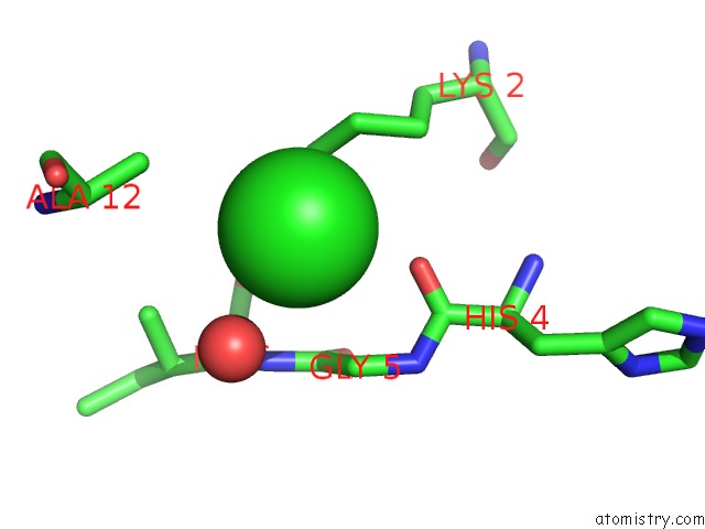
Mono view
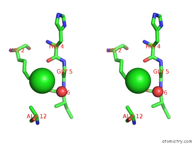
Stereo pair view
Mono view
Stereo pair view
A full contact list of Chlorine with other atoms in the Cl binding
site number 1 of The Structure of Bacillus Subtilis Rbsd Complexed with D-Ribose within 5.0Å range:
|
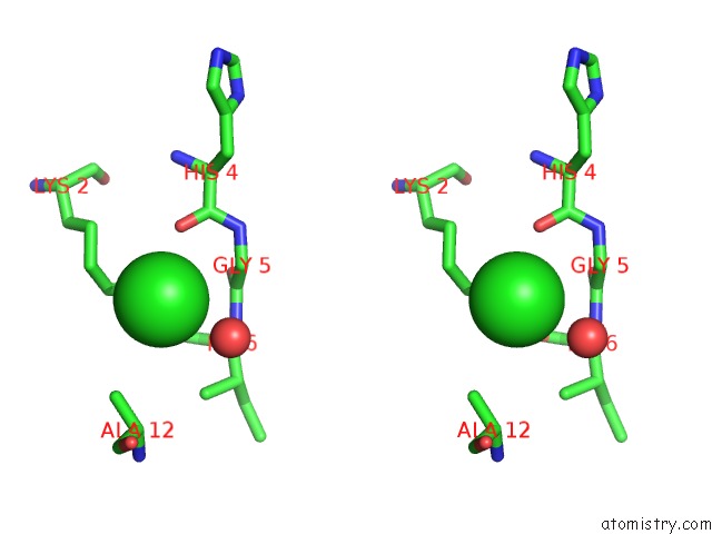
Chlorine binding site 2 out of 2 in 1ogd
Go back to
Chlorine binding site 2 out
of 2 in the The Structure of Bacillus Subtilis Rbsd Complexed with D-Ribose
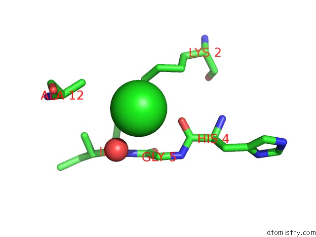
Mono view
Stereo pair view
Mono view
Stereo pair view
A full contact list of Chlorine with other atoms in the Cl binding
site number 2 of The Structure of Bacillus Subtilis Rbsd Complexed with D-Ribose within 5.0Å range:
|
Reference:
M.-S.Kim,
J.Shin,
W.Lee,
H.-S.Lee,
B.-H.Oh.
Crystal Structures of Rbsd Leading to the Identification of Cytoplasmic Sugar-Binding Proteins with A Novel Folding Architecture J.Biol.Chem. V. 278 28173 2003.
ISSN: ISSN 0021-9258
PubMed: 12738765
DOI: 10.1074/JBC.M304523200
Page generated: Sat Jul 20 00:54:36 2024
ISSN: ISSN 0021-9258
PubMed: 12738765
DOI: 10.1074/JBC.M304523200
Last articles
Zn in 9J0NZn in 9J0O
Zn in 9J0P
Zn in 9FJX
Zn in 9EKB
Zn in 9C0F
Zn in 9CAH
Zn in 9CH0
Zn in 9CH3
Zn in 9CH1