Chlorine »
PDB 1on0-1p6y »
1onw »
Chlorine in PDB 1onw: Crystal Structure of Isoaspartyl Dipeptidase From E. Coli
Protein crystallography data
The structure of Crystal Structure of Isoaspartyl Dipeptidase From E. Coli, PDB code: 1onw
was solved by
J.B.Thoden,
R.Marti-Arbona,
F.M.Raushel,
H.M.Holden,
with X-Ray Crystallography technique. A brief refinement statistics is given in the table below:
| Resolution Low / High (Å) | 30.00 / 1.65 |
| Space group | P 4 21 2 |
| Cell size a, b, c (Å), α, β, γ (°) | 116.700, 116.700, 138.500, 90.00, 90.00, 90.00 |
| R / Rfree (%) | n/a / n/a |
Other elements in 1onw:
The structure of Crystal Structure of Isoaspartyl Dipeptidase From E. Coli also contains other interesting chemical elements:
| Magnesium | (Mg) | 1 atom |
| Zinc | (Zn) | 4 atoms |
| Sodium | (Na) | 1 atom |
Chlorine Binding Sites:
The binding sites of Chlorine atom in the Crystal Structure of Isoaspartyl Dipeptidase From E. Coli
(pdb code 1onw). This binding sites where shown within
5.0 Angstroms radius around Chlorine atom.
In total 3 binding sites of Chlorine where determined in the Crystal Structure of Isoaspartyl Dipeptidase From E. Coli, PDB code: 1onw:
Jump to Chlorine binding site number: 1; 2; 3;
In total 3 binding sites of Chlorine where determined in the Crystal Structure of Isoaspartyl Dipeptidase From E. Coli, PDB code: 1onw:
Jump to Chlorine binding site number: 1; 2; 3;
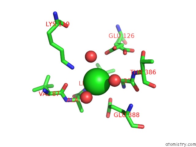
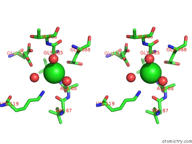
Chlorine binding site 1 out of 3 in 1onw
Go back to
Chlorine binding site 1 out
of 3 in the Crystal Structure of Isoaspartyl Dipeptidase From E. Coli
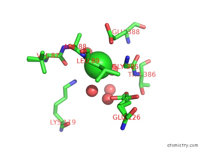
Mono view
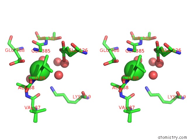
Stereo pair view
Mono view
Stereo pair view
A full contact list of Chlorine with other atoms in the Cl binding
site number 1 of Crystal Structure of Isoaspartyl Dipeptidase From E. Coli within 5.0Å range:
|
Chlorine binding site 2 out of 3 in 1onw
Go back to
Chlorine binding site 2 out
of 3 in the Crystal Structure of Isoaspartyl Dipeptidase From E. Coli
Mono view
Stereo pair view
Mono view
Stereo pair view
A full contact list of Chlorine with other atoms in the Cl binding
site number 2 of Crystal Structure of Isoaspartyl Dipeptidase From E. Coli within 5.0Å range:
|
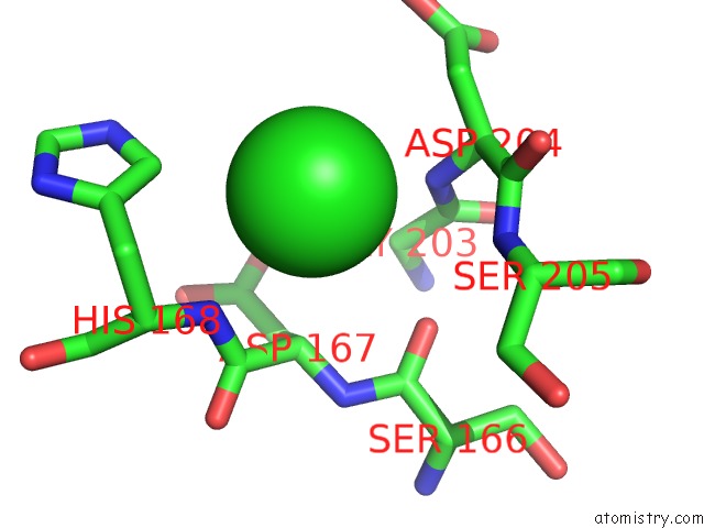
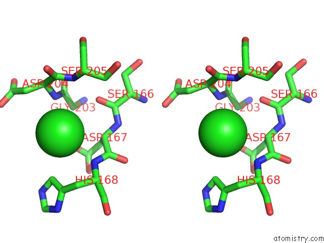
Chlorine binding site 3 out of 3 in 1onw
Go back to
Chlorine binding site 3 out
of 3 in the Crystal Structure of Isoaspartyl Dipeptidase From E. Coli
Mono view
Stereo pair view
Mono view
Stereo pair view
A full contact list of Chlorine with other atoms in the Cl binding
site number 3 of Crystal Structure of Isoaspartyl Dipeptidase From E. Coli within 5.0Å range:
|
Reference:
J.B.Thoden,
R.Marti-Arbona,
F.M.Raushel,
H.M.Holden.
High Resolution X-Ray Structure of Isoaspartyl Dipeptidase From Escherichia Coli Biochemistry V. 42 4874 2003.
ISSN: ISSN 0006-2960
PubMed: 12718528
DOI: 10.1021/BI034233P
Page generated: Sat Jul 20 00:58:40 2024
ISSN: ISSN 0006-2960
PubMed: 12718528
DOI: 10.1021/BI034233P
Last articles
Zn in 9MJ5Zn in 9HNW
Zn in 9G0L
Zn in 9FNE
Zn in 9DZN
Zn in 9E0I
Zn in 9D32
Zn in 9DAK
Zn in 8ZXC
Zn in 8ZUF