Chlorine »
PDB 1qhu-1qul »
1ql7 »
Chlorine in PDB 1ql7: Factor Xa Specific Inhibitor in Complex with Bovine Trypsin
Enzymatic activity of Factor Xa Specific Inhibitor in Complex with Bovine Trypsin
All present enzymatic activity of Factor Xa Specific Inhibitor in Complex with Bovine Trypsin:
3.4.21.4;
3.4.21.4;
Protein crystallography data
The structure of Factor Xa Specific Inhibitor in Complex with Bovine Trypsin, PDB code: 1ql7
was solved by
M.T.Stubbs,
with X-Ray Crystallography technique. A brief refinement statistics is given in the table below:
| Resolution Low / High (Å) | 10.00 / 2.10 |
| Space group | P 21 21 21 |
| Cell size a, b, c (Å), α, β, γ (°) | 54.060, 58.580, 63.140, 90.00, 90.00, 90.00 |
| R / Rfree (%) | 20 / n/a |
Other elements in 1ql7:
The structure of Factor Xa Specific Inhibitor in Complex with Bovine Trypsin also contains other interesting chemical elements:
| Calcium | (Ca) | 1 atom |
Chlorine Binding Sites:
The binding sites of Chlorine atom in the Factor Xa Specific Inhibitor in Complex with Bovine Trypsin
(pdb code 1ql7). This binding sites where shown within
5.0 Angstroms radius around Chlorine atom.
In total only one binding site of Chlorine was determined in the Factor Xa Specific Inhibitor in Complex with Bovine Trypsin, PDB code: 1ql7:
In total only one binding site of Chlorine was determined in the Factor Xa Specific Inhibitor in Complex with Bovine Trypsin, PDB code: 1ql7:
Chlorine binding site 1 out of 1 in 1ql7
Go back to
Chlorine binding site 1 out
of 1 in the Factor Xa Specific Inhibitor in Complex with Bovine Trypsin
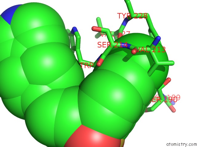
Mono view
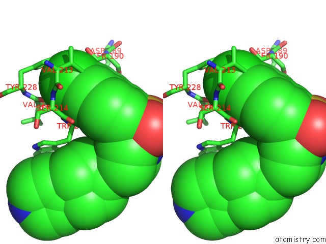
Stereo pair view
Mono view
Stereo pair view
A full contact list of Chlorine with other atoms in the Cl binding
site number 1 of Factor Xa Specific Inhibitor in Complex with Bovine Trypsin within 5.0Å range:
|
Reference:
M.T.Stubbs,
S.Reyda,
F.Dullweber,
M.Moeller,
G.Klebe,
D.Dorsch,
W.W.K.R.Mederski,
H.Wurziger.
pH-Dependent Binding Modes Observed in Trypsin Crystals: Lessons For the Structure-Based Drug Design Chembiochem V. 3 246 2002.
ISSN: ISSN 1439-4227
PubMed: 11921406
DOI: 10.1002/1439-7633(20020301)3:2/3<246::AID-CBIC246>3.0.CO;2-#
Page generated: Sat Jul 20 01:36:33 2024
ISSN: ISSN 1439-4227
PubMed: 11921406
DOI: 10.1002/1439-7633(20020301)3:2/3<246::AID-CBIC246>3.0.CO;2-#
Last articles
Zn in 9JYWZn in 9IR4
Zn in 9IR3
Zn in 9GMX
Zn in 9GMW
Zn in 9JEJ
Zn in 9ERF
Zn in 9ERE
Zn in 9EGV
Zn in 9EGW