Chlorine »
PDB 1qvf-1rkq »
1qz9 »
Chlorine in PDB 1qz9: The Three Dimensional Structure of Kynureninase From Pseudomonas Fluorescens
Enzymatic activity of The Three Dimensional Structure of Kynureninase From Pseudomonas Fluorescens
All present enzymatic activity of The Three Dimensional Structure of Kynureninase From Pseudomonas Fluorescens:
3.7.1.3;
3.7.1.3;
Protein crystallography data
The structure of The Three Dimensional Structure of Kynureninase From Pseudomonas Fluorescens, PDB code: 1qz9
was solved by
C.Momany,
V.Levdikov,
L.Blagova,
S.Lima,
R.S.Phillips,
with X-Ray Crystallography technique. A brief refinement statistics is given in the table below:
| Resolution Low / High (Å) | 19.65 / 1.85 |
| Space group | P 31 2 1 |
| Cell size a, b, c (Å), α, β, γ (°) | 68.076, 68.076, 137.976, 90.00, 90.00, 120.00 |
| R / Rfree (%) | 15.3 / 19.2 |
Chlorine Binding Sites:
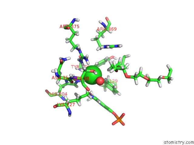
The binding sites of Chlorine atom in the The Three Dimensional Structure of Kynureninase From Pseudomonas Fluorescens
(pdb code 1qz9). This binding sites where shown within
5.0 Angstroms radius around Chlorine atom.
In total only one binding site of Chlorine was determined in the The Three Dimensional Structure of Kynureninase From Pseudomonas Fluorescens, PDB code: 1qz9:
In total only one binding site of Chlorine was determined in the The Three Dimensional Structure of Kynureninase From Pseudomonas Fluorescens, PDB code: 1qz9:
Chlorine binding site 1 out of 1 in 1qz9
Go back to
Chlorine binding site 1 out
of 1 in the The Three Dimensional Structure of Kynureninase From Pseudomonas Fluorescens
Mono view

Stereo pair view
Mono view
Stereo pair view
A full contact list of Chlorine with other atoms in the Cl binding
site number 1 of The Three Dimensional Structure of Kynureninase From Pseudomonas Fluorescens within 5.0Å range:
|
Reference:
C.Momany,
V.Levdikov,
L.Blagova,
S.Lima,
R.S.Phillips.
Three-Dimensional Structure of Kynureninase From Pseudomonas Fluorescens. Biochemistry V. 43 1193 2004.
ISSN: ISSN 0006-2960
PubMed: 14756555
DOI: 10.1021/BI035744E
Page generated: Sat Jul 20 01:47:27 2024
ISSN: ISSN 0006-2960
PubMed: 14756555
DOI: 10.1021/BI035744E
Last articles
Zn in 9J0NZn in 9J0O
Zn in 9J0P
Zn in 9FJX
Zn in 9EKB
Zn in 9C0F
Zn in 9CAH
Zn in 9CH0
Zn in 9CH3
Zn in 9CH1