Chlorine »
PDB 1qvf-1rkq »
1r85 »
Chlorine in PDB 1r85: Crystal Structure of the Extracellular Xylanase From Geobacillus Stearothermophilus T-6 (XT6): the Wt Enzyme (Monoclinic Form) at 1.45A Resolution
Enzymatic activity of Crystal Structure of the Extracellular Xylanase From Geobacillus Stearothermophilus T-6 (XT6): the Wt Enzyme (Monoclinic Form) at 1.45A Resolution
All present enzymatic activity of Crystal Structure of the Extracellular Xylanase From Geobacillus Stearothermophilus T-6 (XT6): the Wt Enzyme (Monoclinic Form) at 1.45A Resolution:
3.2.1.8;
3.2.1.8;
Protein crystallography data
The structure of Crystal Structure of the Extracellular Xylanase From Geobacillus Stearothermophilus T-6 (XT6): the Wt Enzyme (Monoclinic Form) at 1.45A Resolution, PDB code: 1r85
was solved by
M.Bar,
G.Golan,
M.Nechama,
G.Zolotnitsky,
Y.Shoham,
G.Shoham,
with X-Ray Crystallography technique. A brief refinement statistics is given in the table below:
| Resolution Low / High (Å) | 40.00 / 1.45 |
| Space group | C 1 2 1 |
| Cell size a, b, c (Å), α, β, γ (°) | 121.510, 61.710, 89.090, 90.00, 119.07, 90.00 |
| R / Rfree (%) | 12.6 / 18.1 |
Other elements in 1r85:
The structure of Crystal Structure of the Extracellular Xylanase From Geobacillus Stearothermophilus T-6 (XT6): the Wt Enzyme (Monoclinic Form) at 1.45A Resolution also contains other interesting chemical elements:
| Zinc | (Zn) | 7 atoms |
Chlorine Binding Sites:
The binding sites of Chlorine atom in the Crystal Structure of the Extracellular Xylanase From Geobacillus Stearothermophilus T-6 (XT6): the Wt Enzyme (Monoclinic Form) at 1.45A Resolution
(pdb code 1r85). This binding sites where shown within
5.0 Angstroms radius around Chlorine atom.
In total 2 binding sites of Chlorine where determined in the Crystal Structure of the Extracellular Xylanase From Geobacillus Stearothermophilus T-6 (XT6): the Wt Enzyme (Monoclinic Form) at 1.45A Resolution, PDB code: 1r85:
Jump to Chlorine binding site number: 1; 2;
In total 2 binding sites of Chlorine where determined in the Crystal Structure of the Extracellular Xylanase From Geobacillus Stearothermophilus T-6 (XT6): the Wt Enzyme (Monoclinic Form) at 1.45A Resolution, PDB code: 1r85:
Jump to Chlorine binding site number: 1; 2;
Chlorine binding site 1 out of 2 in 1r85
Go back to
Chlorine binding site 1 out
of 2 in the Crystal Structure of the Extracellular Xylanase From Geobacillus Stearothermophilus T-6 (XT6): the Wt Enzyme (Monoclinic Form) at 1.45A Resolution
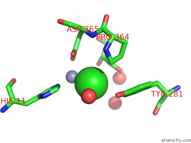
Mono view
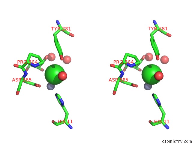
Stereo pair view
Mono view
Stereo pair view
A full contact list of Chlorine with other atoms in the Cl binding
site number 1 of Crystal Structure of the Extracellular Xylanase From Geobacillus Stearothermophilus T-6 (XT6): the Wt Enzyme (Monoclinic Form) at 1.45A Resolution within 5.0Å range:
|
Chlorine binding site 2 out of 2 in 1r85
Go back to
Chlorine binding site 2 out
of 2 in the Crystal Structure of the Extracellular Xylanase From Geobacillus Stearothermophilus T-6 (XT6): the Wt Enzyme (Monoclinic Form) at 1.45A Resolution
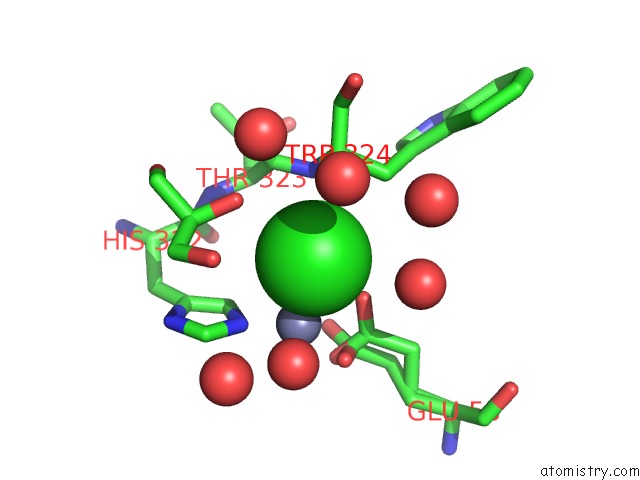
Mono view
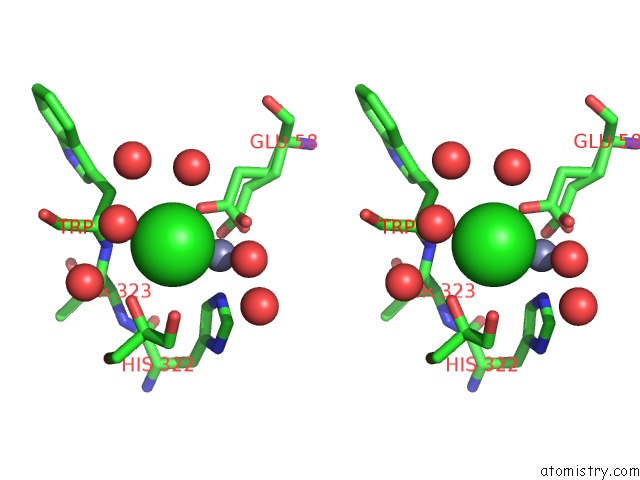
Stereo pair view
Mono view
Stereo pair view
A full contact list of Chlorine with other atoms in the Cl binding
site number 2 of Crystal Structure of the Extracellular Xylanase From Geobacillus Stearothermophilus T-6 (XT6): the Wt Enzyme (Monoclinic Form) at 1.45A Resolution within 5.0Å range:
|
Reference:
G.Zolotnitsky,
U.Cogan,
N.Adir,
V.Solomon,
G.Shoham,
Y.Shoham.
Mapping Glycoside Hydrolase Substrate Subsites By Isothermal Titration Calorimetry. Proc.Natl.Acad.Sci.Usa V. 101 11275 2004.
ISSN: ISSN 0027-8424
PubMed: 15277671
DOI: 10.1073/PNAS.0404311101
Page generated: Sat Jul 20 01:51:05 2024
ISSN: ISSN 0027-8424
PubMed: 15277671
DOI: 10.1073/PNAS.0404311101
Last articles
Zn in 9J0NZn in 9J0O
Zn in 9J0P
Zn in 9FJX
Zn in 9EKB
Zn in 9C0F
Zn in 9CAH
Zn in 9CH0
Zn in 9CH3
Zn in 9CH1