Chlorine »
PDB 1rky-1s8f »
1s36 »
Chlorine in PDB 1s36: Crystal Structure of A CA2+-Discharged Photoprotein: Implications For the Mechanisms of the Calcium Trigger and the Bioluminescence
Protein crystallography data
The structure of Crystal Structure of A CA2+-Discharged Photoprotein: Implications For the Mechanisms of the Calcium Trigger and the Bioluminescence, PDB code: 1s36
was solved by
L.Deng,
S.V.Markova,
E.S.Vysotski,
Z.-J.Liu,
J.Lee,
J.Rose,
B.-C.Wang,
Southeast Collaboratory For Structural Genomics (Secsg),
with X-Ray Crystallography technique. A brief refinement statistics is given in the table below:
| Resolution Low / High (Å) | 50.00 / 1.96 |
| Space group | P 41 21 2 |
| Cell size a, b, c (Å), α, β, γ (°) | 53.445, 53.445, 144.032, 90.00, 90.00, 90.00 |
| R / Rfree (%) | 22.3 / 25.9 |
Other elements in 1s36:
The structure of Crystal Structure of A CA2+-Discharged Photoprotein: Implications For the Mechanisms of the Calcium Trigger and the Bioluminescence also contains other interesting chemical elements:
| Sodium | (Na) | 1 atom |
Chlorine Binding Sites:
The binding sites of Chlorine atom in the Crystal Structure of A CA2+-Discharged Photoprotein: Implications For the Mechanisms of the Calcium Trigger and the Bioluminescence
(pdb code 1s36). This binding sites where shown within
5.0 Angstroms radius around Chlorine atom.
In total only one binding site of Chlorine was determined in the Crystal Structure of A CA2+-Discharged Photoprotein: Implications For the Mechanisms of the Calcium Trigger and the Bioluminescence, PDB code: 1s36:
In total only one binding site of Chlorine was determined in the Crystal Structure of A CA2+-Discharged Photoprotein: Implications For the Mechanisms of the Calcium Trigger and the Bioluminescence, PDB code: 1s36:
Chlorine binding site 1 out of 1 in 1s36
Go back to
Chlorine binding site 1 out
of 1 in the Crystal Structure of A CA2+-Discharged Photoprotein: Implications For the Mechanisms of the Calcium Trigger and the Bioluminescence
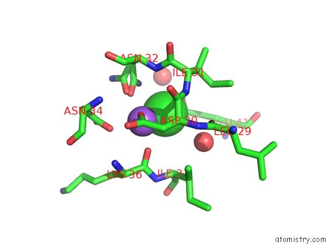
Mono view
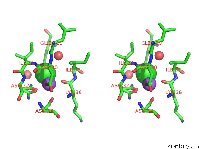
Stereo pair view
Mono view
Stereo pair view
A full contact list of Chlorine with other atoms in the Cl binding
site number 1 of Crystal Structure of A CA2+-Discharged Photoprotein: Implications For the Mechanisms of the Calcium Trigger and the Bioluminescence within 5.0Å range:
|
Reference:
L.Deng,
S.V.Markova,
E.S.Vysotski,
Z.-J.Liu,
J.Lee,
J.Rose,
B.-C.Wang.
Crystal Structure of A CA2+-Discharged Photoprotein: Implications For Mechanisms of the Calcium Trigger and Bioluminescence J.Biol.Chem. V. 279 33647 2004.
ISSN: ISSN 0021-9258
PubMed: 15155735
DOI: 10.1074/JBC.M402427200
Page generated: Sat Jul 20 02:02:25 2024
ISSN: ISSN 0021-9258
PubMed: 15155735
DOI: 10.1074/JBC.M402427200
Last articles
Zn in 9J0NZn in 9J0O
Zn in 9J0P
Zn in 9FJX
Zn in 9EKB
Zn in 9C0F
Zn in 9CAH
Zn in 9CH0
Zn in 9CH3
Zn in 9CH1