Chlorine »
PDB 1rky-1s8f »
1s4i »
Chlorine in PDB 1s4i: Crystal Structure of A Sod-Like Protein From Bacillus Subtilis
Enzymatic activity of Crystal Structure of A Sod-Like Protein From Bacillus Subtilis
All present enzymatic activity of Crystal Structure of A Sod-Like Protein From Bacillus Subtilis:
1.15.1.1;
1.15.1.1;
Protein crystallography data
The structure of Crystal Structure of A Sod-Like Protein From Bacillus Subtilis, PDB code: 1s4i
was solved by
L.Banci,
I.Bertini,
V.Calderone,
F.Cramaro,
R.Del Conte,
A.Fantoni,
S.Mangani,
A.Quattrone,
M.S.Viezzoli,
with X-Ray Crystallography technique. A brief refinement statistics is given in the table below:
| Resolution Low / High (Å) | 37.01 / 1.80 |
| Space group | P 1 |
| Cell size a, b, c (Å), α, β, γ (°) | 38.225, 61.108, 64.915, 84.35, 76.02, 90.42 |
| R / Rfree (%) | 22.1 / 25.8 |
Other elements in 1s4i:
The structure of Crystal Structure of A Sod-Like Protein From Bacillus Subtilis also contains other interesting chemical elements:
| Zinc | (Zn) | 6 atoms |
Chlorine Binding Sites:
The binding sites of Chlorine atom in the Crystal Structure of A Sod-Like Protein From Bacillus Subtilis
(pdb code 1s4i). This binding sites where shown within
5.0 Angstroms radius around Chlorine atom.
In total 4 binding sites of Chlorine where determined in the Crystal Structure of A Sod-Like Protein From Bacillus Subtilis, PDB code: 1s4i:
Jump to Chlorine binding site number: 1; 2; 3; 4;
In total 4 binding sites of Chlorine where determined in the Crystal Structure of A Sod-Like Protein From Bacillus Subtilis, PDB code: 1s4i:
Jump to Chlorine binding site number: 1; 2; 3; 4;
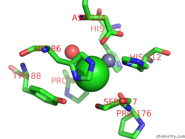
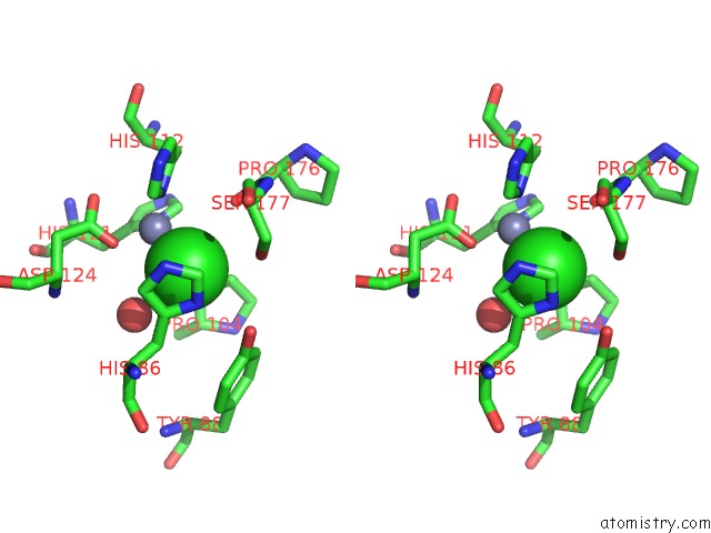
Chlorine binding site 1 out of 4 in 1s4i
Go back to
Chlorine binding site 1 out
of 4 in the Crystal Structure of A Sod-Like Protein From Bacillus Subtilis
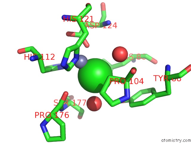
Mono view
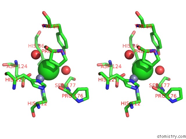
Stereo pair view
Mono view
Stereo pair view
A full contact list of Chlorine with other atoms in the Cl binding
site number 1 of Crystal Structure of A Sod-Like Protein From Bacillus Subtilis within 5.0Å range:
|
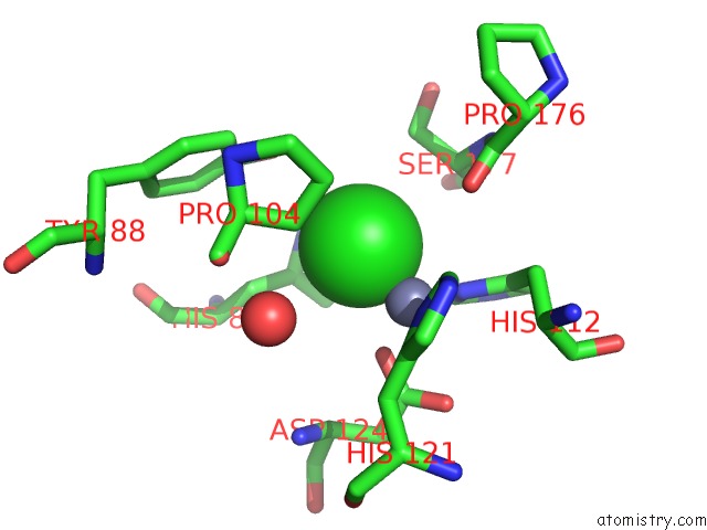
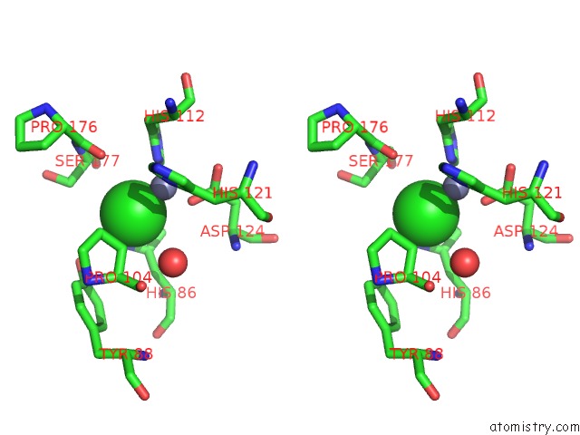
Chlorine binding site 2 out of 4 in 1s4i
Go back to
Chlorine binding site 2 out
of 4 in the Crystal Structure of A Sod-Like Protein From Bacillus Subtilis
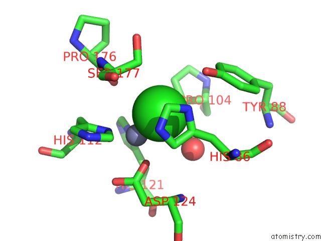
Mono view
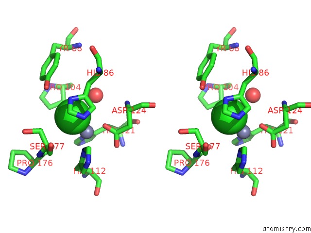
Stereo pair view
Mono view
Stereo pair view
A full contact list of Chlorine with other atoms in the Cl binding
site number 2 of Crystal Structure of A Sod-Like Protein From Bacillus Subtilis within 5.0Å range:
|
Chlorine binding site 3 out of 4 in 1s4i
Go back to
Chlorine binding site 3 out
of 4 in the Crystal Structure of A Sod-Like Protein From Bacillus Subtilis
Mono view
Stereo pair view
Mono view
Stereo pair view
A full contact list of Chlorine with other atoms in the Cl binding
site number 3 of Crystal Structure of A Sod-Like Protein From Bacillus Subtilis within 5.0Å range:
|
Chlorine binding site 4 out of 4 in 1s4i
Go back to
Chlorine binding site 4 out
of 4 in the Crystal Structure of A Sod-Like Protein From Bacillus Subtilis
Mono view
Stereo pair view
Mono view
Stereo pair view
A full contact list of Chlorine with other atoms in the Cl binding
site number 4 of Crystal Structure of A Sod-Like Protein From Bacillus Subtilis within 5.0Å range:
|
Reference:
L.Banci,
I.Bertini,
V.Calderone,
F.Cramaro,
R.Del Conte,
A.Fantoni,
S.Mangani,
A.Quattrone,
M.S.Viezzoli.
A Prokaryotic Superoxide Dismutase Paralog Lacking Two Cu Ligands: From Largely Unstructured in Solution to Ordered in the Crystal. Proc.Natl.Acad.Sci.Usa V. 102 7541 2005.
ISSN: ISSN 0027-8424
PubMed: 15897454
DOI: 10.1073/PNAS.0502450102
Page generated: Sat Jul 20 02:02:33 2024
ISSN: ISSN 0027-8424
PubMed: 15897454
DOI: 10.1073/PNAS.0502450102
Last articles
Zn in 9J0NZn in 9J0O
Zn in 9J0P
Zn in 9FJX
Zn in 9EKB
Zn in 9C0F
Zn in 9CAH
Zn in 9CH0
Zn in 9CH3
Zn in 9CH1