Chlorine »
PDB 1s9e-1swy »
1suo »
Chlorine in PDB 1suo: Structure of Mammalian Cytochrome P450 2B4 with Bound 4-(4- Chlorophenyl)Imidazole
Enzymatic activity of Structure of Mammalian Cytochrome P450 2B4 with Bound 4-(4- Chlorophenyl)Imidazole
All present enzymatic activity of Structure of Mammalian Cytochrome P450 2B4 with Bound 4-(4- Chlorophenyl)Imidazole:
1.14.14.1;
1.14.14.1;
Protein crystallography data
The structure of Structure of Mammalian Cytochrome P450 2B4 with Bound 4-(4- Chlorophenyl)Imidazole, PDB code: 1suo
was solved by
E.E.Scott,
M.A.White,
Y.A.He,
E.F.Johnson,
C.D.Stout,
J.R.Halpert,
with X-Ray Crystallography technique. A brief refinement statistics is given in the table below:
| Resolution Low / High (Å) | 49.00 / 1.90 |
| Space group | P 63 2 2 |
| Cell size a, b, c (Å), α, β, γ (°) | 233.386, 233.386, 56.380, 90.00, 90.00, 120.00 |
| R / Rfree (%) | 21.5 / 23.9 |
Other elements in 1suo:
The structure of Structure of Mammalian Cytochrome P450 2B4 with Bound 4-(4- Chlorophenyl)Imidazole also contains other interesting chemical elements:
| Iron | (Fe) | 1 atom |
Chlorine Binding Sites:
The binding sites of Chlorine atom in the Structure of Mammalian Cytochrome P450 2B4 with Bound 4-(4- Chlorophenyl)Imidazole
(pdb code 1suo). This binding sites where shown within
5.0 Angstroms radius around Chlorine atom.
In total only one binding site of Chlorine was determined in the Structure of Mammalian Cytochrome P450 2B4 with Bound 4-(4- Chlorophenyl)Imidazole, PDB code: 1suo:
In total only one binding site of Chlorine was determined in the Structure of Mammalian Cytochrome P450 2B4 with Bound 4-(4- Chlorophenyl)Imidazole, PDB code: 1suo:
Chlorine binding site 1 out of 1 in 1suo
Go back to
Chlorine binding site 1 out
of 1 in the Structure of Mammalian Cytochrome P450 2B4 with Bound 4-(4- Chlorophenyl)Imidazole
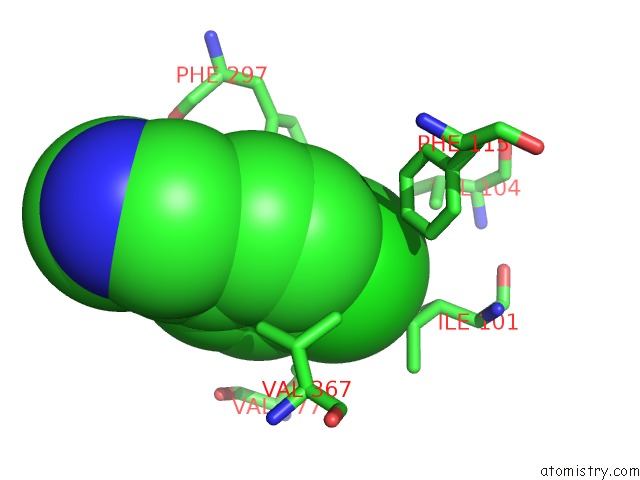
Mono view
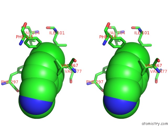
Stereo pair view
Mono view
Stereo pair view
A full contact list of Chlorine with other atoms in the Cl binding
site number 1 of Structure of Mammalian Cytochrome P450 2B4 with Bound 4-(4- Chlorophenyl)Imidazole within 5.0Å range:
|
Reference:
E.E.Scott,
M.A.White,
Y.A.He,
E.F.Johnson,
C.D.Stout,
J.R.Halpert.
Structure of Mammalian Cytochrome P450 2B4 Complexed with 4-(4-Chlorophenyl)Imidazole at 1.9 {Angstrom} Resolution: Insight Into the Range of P450 Conformations and Coordination of Redox Partner Binding. J.Biol.Chem. V. 279 27294 2004.
ISSN: ISSN 0021-9258
PubMed: 15100217
DOI: 10.1074/JBC.M403349200
Page generated: Sat Jul 20 02:15:31 2024
ISSN: ISSN 0021-9258
PubMed: 15100217
DOI: 10.1074/JBC.M403349200
Last articles
Zn in 9J0NZn in 9J0O
Zn in 9J0P
Zn in 9FJX
Zn in 9EKB
Zn in 9C0F
Zn in 9CAH
Zn in 9CH0
Zn in 9CH3
Zn in 9CH1