Chlorine »
PDB 1ti7-1u33 »
1tpk »
Chlorine in PDB 1tpk: Crystal Structure of the Kringle-2 Domain of Tissue Plasminogen Activator at 2.4-Angstroms Resolution
Enzymatic activity of Crystal Structure of the Kringle-2 Domain of Tissue Plasminogen Activator at 2.4-Angstroms Resolution
All present enzymatic activity of Crystal Structure of the Kringle-2 Domain of Tissue Plasminogen Activator at 2.4-Angstroms Resolution:
3.4.21.31;
3.4.21.31;
Protein crystallography data
The structure of Crystal Structure of the Kringle-2 Domain of Tissue Plasminogen Activator at 2.4-Angstroms Resolution, PDB code: 1tpk
was solved by
A.M.De Vos,
M.H.Ultsch,
R.F.Kelley,
K.Padmanabhan,
A.Tulinsky,
M.L.Westbrook,
A.A.Kossiakoff,
with X-Ray Crystallography technique. A brief refinement statistics is given in the table below:
| Resolution Low / High (Å) | 10.00 / 2.40 |
| Space group | P 1 21 1 |
| Cell size a, b, c (Å), α, β, γ (°) | 54.800, 63.580, 46.580, 90.00, 106.73, 90.00 |
| R / Rfree (%) | 18.4 / n/a |
Chlorine Binding Sites:
The binding sites of Chlorine atom in the Crystal Structure of the Kringle-2 Domain of Tissue Plasminogen Activator at 2.4-Angstroms Resolution
(pdb code 1tpk). This binding sites where shown within
5.0 Angstroms radius around Chlorine atom.
In total 3 binding sites of Chlorine where determined in the Crystal Structure of the Kringle-2 Domain of Tissue Plasminogen Activator at 2.4-Angstroms Resolution, PDB code: 1tpk:
Jump to Chlorine binding site number: 1; 2; 3;
In total 3 binding sites of Chlorine where determined in the Crystal Structure of the Kringle-2 Domain of Tissue Plasminogen Activator at 2.4-Angstroms Resolution, PDB code: 1tpk:
Jump to Chlorine binding site number: 1; 2; 3;
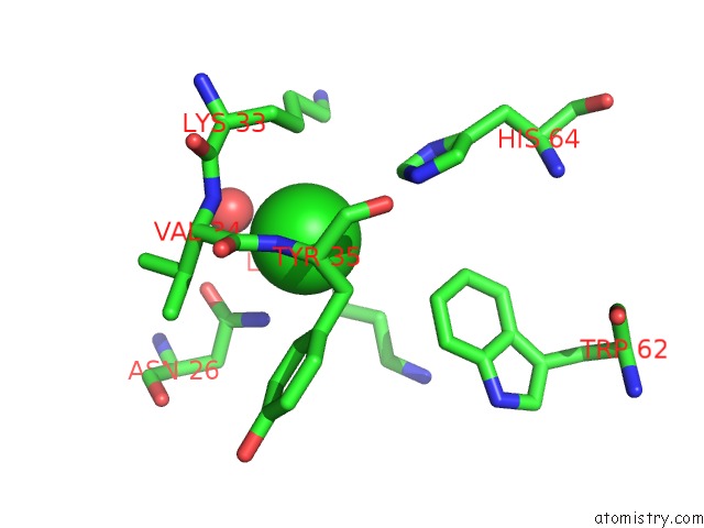
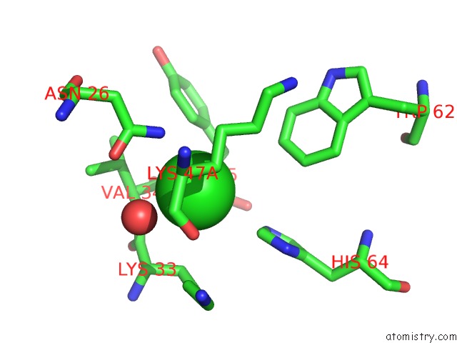
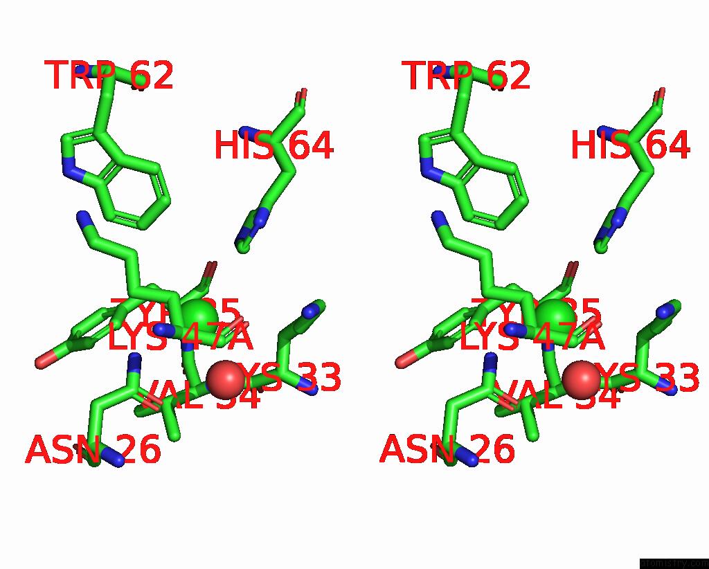
Chlorine binding site 1 out of 3 in 1tpk
Go back to
Chlorine binding site 1 out
of 3 in the Crystal Structure of the Kringle-2 Domain of Tissue Plasminogen Activator at 2.4-Angstroms Resolution
Mono view
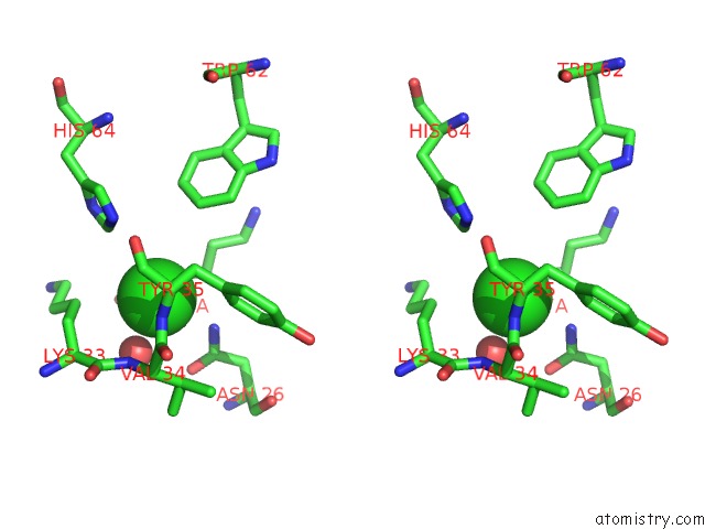
Stereo pair view
Mono view
Stereo pair view
A full contact list of Chlorine with other atoms in the Cl binding
site number 1 of Crystal Structure of the Kringle-2 Domain of Tissue Plasminogen Activator at 2.4-Angstroms Resolution within 5.0Å range:
|
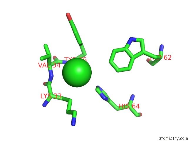
Chlorine binding site 2 out of 3 in 1tpk
Go back to
Chlorine binding site 2 out
of 3 in the Crystal Structure of the Kringle-2 Domain of Tissue Plasminogen Activator at 2.4-Angstroms Resolution
Mono view
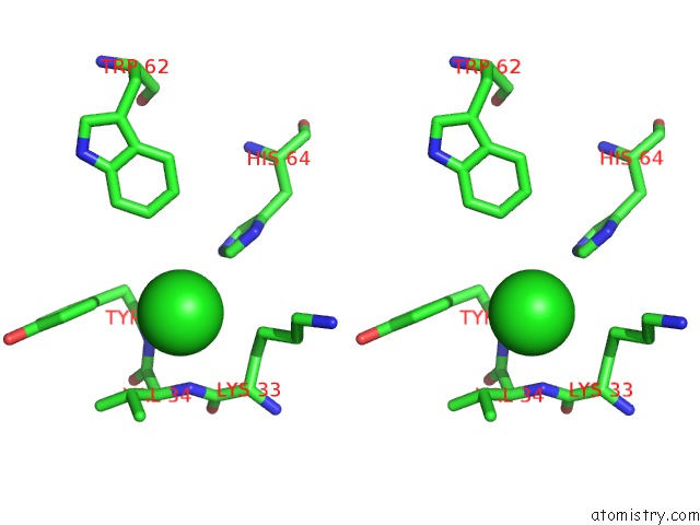
Stereo pair view
Mono view
Stereo pair view
A full contact list of Chlorine with other atoms in the Cl binding
site number 2 of Crystal Structure of the Kringle-2 Domain of Tissue Plasminogen Activator at 2.4-Angstroms Resolution within 5.0Å range:
|
Chlorine binding site 3 out of 3 in 1tpk
Go back to
Chlorine binding site 3 out
of 3 in the Crystal Structure of the Kringle-2 Domain of Tissue Plasminogen Activator at 2.4-Angstroms Resolution
Mono view
Stereo pair view
Mono view
Stereo pair view
A full contact list of Chlorine with other atoms in the Cl binding
site number 3 of Crystal Structure of the Kringle-2 Domain of Tissue Plasminogen Activator at 2.4-Angstroms Resolution within 5.0Å range:
|
Reference:
A.M.De Vos,
M.H.Ultsch,
R.F.Kelley,
K.Padmanabhan,
A.Tulinsky,
M.L.Westbrook,
A.A.Kossiakoff.
Crystal Structure of the Kringle 2 Domain of Tissue Plasminogen Activator at 2.4-A Resolution. Biochemistry V. 31 270 1992.
ISSN: ISSN 0006-2960
PubMed: 1310033
DOI: 10.1021/BI00116A037
Page generated: Sat Jul 20 02:35:19 2024
ISSN: ISSN 0006-2960
PubMed: 1310033
DOI: 10.1021/BI00116A037
Last articles
Zn in 9J0NZn in 9J0O
Zn in 9J0P
Zn in 9FJX
Zn in 9EKB
Zn in 9C0F
Zn in 9CAH
Zn in 9CH0
Zn in 9CH3
Zn in 9CH1