Chlorine »
PDB 1v8o-1vq4 »
1va0 »
Chlorine in PDB 1va0: Crystal Structure of the Native Form of Uroporphyrin III C-Methyl Transferase From Thermus Thermophilus
Enzymatic activity of Crystal Structure of the Native Form of Uroporphyrin III C-Methyl Transferase From Thermus Thermophilus
All present enzymatic activity of Crystal Structure of the Native Form of Uroporphyrin III C-Methyl Transferase From Thermus Thermophilus:
2.1.1.107;
2.1.1.107;
Protein crystallography data
The structure of Crystal Structure of the Native Form of Uroporphyrin III C-Methyl Transferase From Thermus Thermophilus, PDB code: 1va0
was solved by
P.H.Rehse,
T.Kitao,
T.H.Tahirov,
Riken Structural Genomics/Proteomicsinitiative (Rsgi),
with X-Ray Crystallography technique. A brief refinement statistics is given in the table below:
| Resolution Low / High (Å) | 31.78 / 1.97 |
| Space group | P 21 21 21 |
| Cell size a, b, c (Å), α, β, γ (°) | 60.500, 63.568, 131.902, 90.00, 90.00, 90.00 |
| R / Rfree (%) | 19.9 / 22.7 |
Chlorine Binding Sites:
The binding sites of Chlorine atom in the Crystal Structure of the Native Form of Uroporphyrin III C-Methyl Transferase From Thermus Thermophilus
(pdb code 1va0). This binding sites where shown within
5.0 Angstroms radius around Chlorine atom.
In total 3 binding sites of Chlorine where determined in the Crystal Structure of the Native Form of Uroporphyrin III C-Methyl Transferase From Thermus Thermophilus, PDB code: 1va0:
Jump to Chlorine binding site number: 1; 2; 3;
In total 3 binding sites of Chlorine where determined in the Crystal Structure of the Native Form of Uroporphyrin III C-Methyl Transferase From Thermus Thermophilus, PDB code: 1va0:
Jump to Chlorine binding site number: 1; 2; 3;
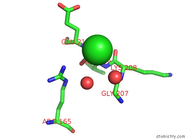
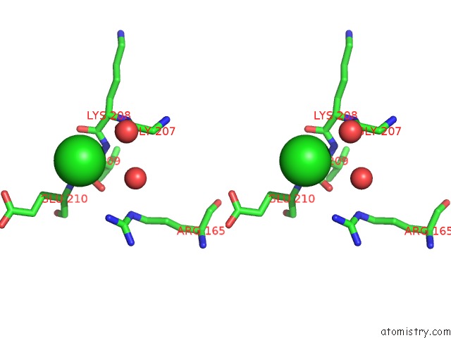
Chlorine binding site 1 out of 3 in 1va0
Go back to
Chlorine binding site 1 out
of 3 in the Crystal Structure of the Native Form of Uroporphyrin III C-Methyl Transferase From Thermus Thermophilus
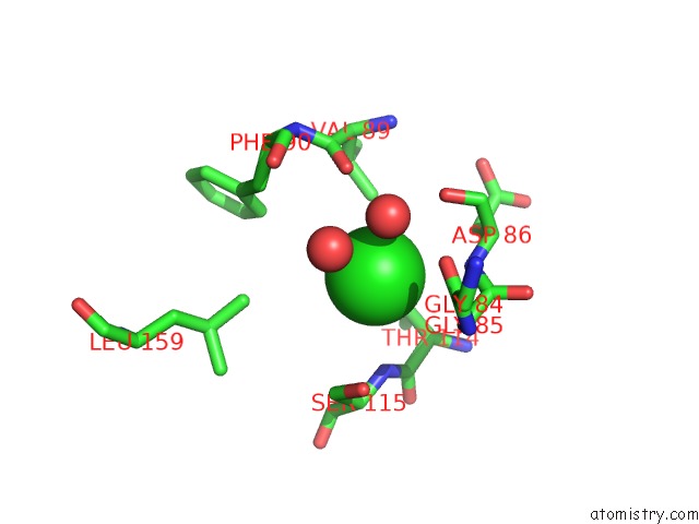
Mono view
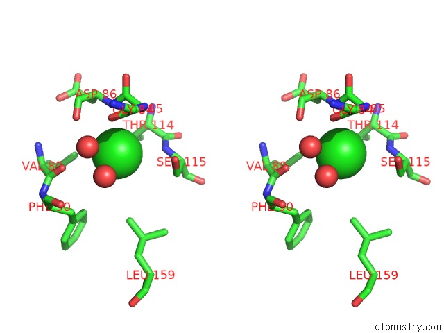
Stereo pair view
Mono view
Stereo pair view
A full contact list of Chlorine with other atoms in the Cl binding
site number 1 of Crystal Structure of the Native Form of Uroporphyrin III C-Methyl Transferase From Thermus Thermophilus within 5.0Å range:
|
Chlorine binding site 2 out of 3 in 1va0
Go back to
Chlorine binding site 2 out
of 3 in the Crystal Structure of the Native Form of Uroporphyrin III C-Methyl Transferase From Thermus Thermophilus
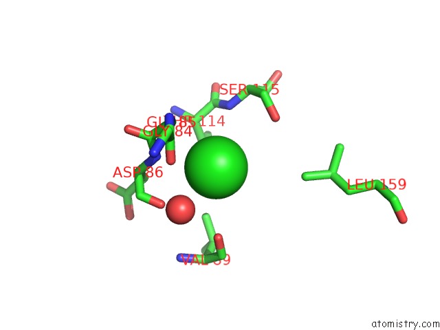
Mono view
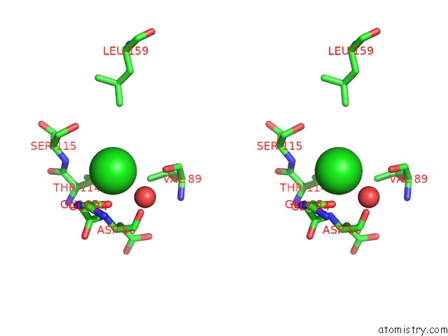
Stereo pair view
Mono view
Stereo pair view
A full contact list of Chlorine with other atoms in the Cl binding
site number 2 of Crystal Structure of the Native Form of Uroporphyrin III C-Methyl Transferase From Thermus Thermophilus within 5.0Å range:
|
Chlorine binding site 3 out of 3 in 1va0
Go back to
Chlorine binding site 3 out
of 3 in the Crystal Structure of the Native Form of Uroporphyrin III C-Methyl Transferase From Thermus Thermophilus
Mono view
Stereo pair view
Mono view
Stereo pair view
A full contact list of Chlorine with other atoms in the Cl binding
site number 3 of Crystal Structure of the Native Form of Uroporphyrin III C-Methyl Transferase From Thermus Thermophilus within 5.0Å range:
|
Reference:
P.H.Rehse,
T.Kitao,
T.H.Tahirov.
Structure of A Closed-Form Uroporphyrinogen-III C-Methyltransferase From Thermus Thermophilus. Acta Crystallogr.,Sect.D V. 61 913 2005.
ISSN: ISSN 0907-4449
PubMed: 15983414
DOI: 10.1107/S0907444905008838
Page generated: Sat Jul 20 03:00:02 2024
ISSN: ISSN 0907-4449
PubMed: 15983414
DOI: 10.1107/S0907444905008838
Last articles
Zn in 9J0NZn in 9J0O
Zn in 9J0P
Zn in 9FJX
Zn in 9EKB
Zn in 9C0F
Zn in 9CAH
Zn in 9CH0
Zn in 9CH3
Zn in 9CH1