Chlorine »
PDB 1v8o-1vq4 »
1vg0 »
Chlorine in PDB 1vg0: The Crystal Structures of the Rep-1 Protein in Complex with Monoprenylated RAB7 Protein
Protein crystallography data
The structure of The Crystal Structures of the Rep-1 Protein in Complex with Monoprenylated RAB7 Protein, PDB code: 1vg0
was solved by
A.Rak,
O.Pylypenko,
A.Niculae,
K.Pyatkov,
R.S.Goody,
K.Alexandrov,
with X-Ray Crystallography technique. A brief refinement statistics is given in the table below:
| Resolution Low / High (Å) | 18.00 / 2.20 |
| Space group | P 21 21 2 |
| Cell size a, b, c (Å), α, β, γ (°) | 64.300, 105.300, 132.600, 90.00, 90.00, 90.00 |
| R / Rfree (%) | 18.8 / 23.3 |
Other elements in 1vg0:
The structure of The Crystal Structures of the Rep-1 Protein in Complex with Monoprenylated RAB7 Protein also contains other interesting chemical elements:
| Magnesium | (Mg) | 1 atom |
Chlorine Binding Sites:
The binding sites of Chlorine atom in the The Crystal Structures of the Rep-1 Protein in Complex with Monoprenylated RAB7 Protein
(pdb code 1vg0). This binding sites where shown within
5.0 Angstroms radius around Chlorine atom.
In total only one binding site of Chlorine was determined in the The Crystal Structures of the Rep-1 Protein in Complex with Monoprenylated RAB7 Protein, PDB code: 1vg0:
In total only one binding site of Chlorine was determined in the The Crystal Structures of the Rep-1 Protein in Complex with Monoprenylated RAB7 Protein, PDB code: 1vg0:
Chlorine binding site 1 out of 1 in 1vg0
Go back to
Chlorine binding site 1 out
of 1 in the The Crystal Structures of the Rep-1 Protein in Complex with Monoprenylated RAB7 Protein
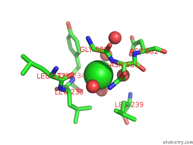
Mono view
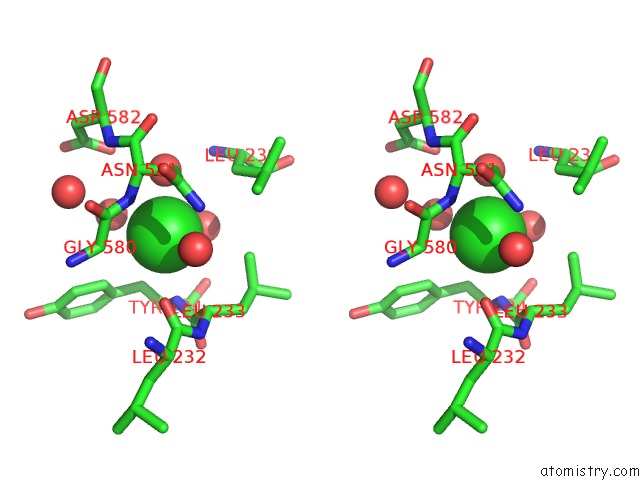
Stereo pair view
Mono view
Stereo pair view
A full contact list of Chlorine with other atoms in the Cl binding
site number 1 of The Crystal Structures of the Rep-1 Protein in Complex with Monoprenylated RAB7 Protein within 5.0Å range:
|
Reference:
A.Rak,
O.Pylypenko,
A.Niculae,
K.Pyatkov,
R.S.Goody,
K.Alexandrov.
Structure of the RAB7:Rep-1 Complex: Insights Into the Mechanism of Rab Prenylation and Choroideremia Disease Cell(Cambridge,Mass.) V. 117 749 2004.
ISSN: ISSN 0092-8674
PubMed: 15186776
DOI: 10.1016/J.CELL.2004.05.017
Page generated: Sat Jul 20 03:03:02 2024
ISSN: ISSN 0092-8674
PubMed: 15186776
DOI: 10.1016/J.CELL.2004.05.017
Last articles
Zn in 9J0NZn in 9J0O
Zn in 9J0P
Zn in 9FJX
Zn in 9EKB
Zn in 9C0F
Zn in 9CAH
Zn in 9CH0
Zn in 9CH3
Zn in 9CH1