Chlorine »
PDB 1v8o-1vq4 »
1vk3 »
Chlorine in PDB 1vk3: Crystal Structure of Phosphoribosylformylglycinamidine Synthase II (TM1246) From Thermotoga Maritima at 2.15 A Resolution
Enzymatic activity of Crystal Structure of Phosphoribosylformylglycinamidine Synthase II (TM1246) From Thermotoga Maritima at 2.15 A Resolution
All present enzymatic activity of Crystal Structure of Phosphoribosylformylglycinamidine Synthase II (TM1246) From Thermotoga Maritima at 2.15 A Resolution:
6.3.5.3;
6.3.5.3;
Protein crystallography data
The structure of Crystal Structure of Phosphoribosylformylglycinamidine Synthase II (TM1246) From Thermotoga Maritima at 2.15 A Resolution, PDB code: 1vk3
was solved by
Joint Center For Structural Genomics (Jcsg),
with X-Ray Crystallography technique. A brief refinement statistics is given in the table below:
| Resolution Low / High (Å) | 63.26 / 2.15 |
| Space group | P 21 21 21 |
| Cell size a, b, c (Å), α, β, γ (°) | 59.775, 72.689, 128.418, 90.00, 90.00, 90.00 |
| R / Rfree (%) | 18.6 / 25.4 |
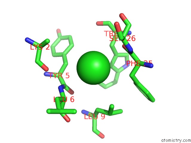
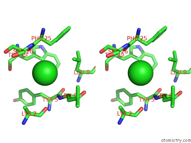
Chlorine Binding Sites:
The binding sites of Chlorine atom in the Crystal Structure of Phosphoribosylformylglycinamidine Synthase II (TM1246) From Thermotoga Maritima at 2.15 A Resolution
(pdb code 1vk3). This binding sites where shown within
5.0 Angstroms radius around Chlorine atom.
In total only one binding site of Chlorine was determined in the Crystal Structure of Phosphoribosylformylglycinamidine Synthase II (TM1246) From Thermotoga Maritima at 2.15 A Resolution, PDB code: 1vk3:
In total only one binding site of Chlorine was determined in the Crystal Structure of Phosphoribosylformylglycinamidine Synthase II (TM1246) From Thermotoga Maritima at 2.15 A Resolution, PDB code: 1vk3:
Chlorine binding site 1 out of 1 in 1vk3
Go back to
Chlorine binding site 1 out
of 1 in the Crystal Structure of Phosphoribosylformylglycinamidine Synthase II (TM1246) From Thermotoga Maritima at 2.15 A Resolution
Mono view
Stereo pair view
Mono view
Stereo pair view
A full contact list of Chlorine with other atoms in the Cl binding
site number 1 of Crystal Structure of Phosphoribosylformylglycinamidine Synthase II (TM1246) From Thermotoga Maritima at 2.15 A Resolution within 5.0Å range:
|
Reference:
I.I.Mathews,
S.S.Krishna,
R.Schwarzenbacher,
D.Mcmullan,
P.Abdubek,
E.Ambing,
J.M.Canaves,
H.J.Chiu,
A.M.Deacon,
M.Didonato,
M.A.Elsliger,
A.Godzik,
C.Grittini,
S.K.Grzechnik,
J.Hale,
E.Hampton,
G.W.Han,
J.Haugen,
L.Jaroszewski,
H.E.Klock,
E.Koesema,
A.Kreusch,
P.Kuhn,
S.A.Lesley,
I.Levin,
M.D.Miller,
K.Moy,
E.Nigoghossian,
J.Paulsen,
K.Quijano,
R.Reyes,
G.Spraggon,
R.C.Stevens,
H.Van Den Bedem,
J.Velasquez,
A.White,
G.Wolf,
Q.Xu,
K.O.Hodgson,
J.Wooley,
I.A.Wilson.
Crystal Structure of Phosphoribosylformylglycinamidine Synthase II (Smpurl) From Thermotoga Maritima at 2.15 A Resolution. Proteins V. 63 1106 2006.
ISSN: ISSN 0887-3585
PubMed: 16544324
DOI: 10.1002/PROT.20650
Page generated: Sat Jul 20 03:04:13 2024
ISSN: ISSN 0887-3585
PubMed: 16544324
DOI: 10.1002/PROT.20650
Last articles
Zn in 9J0NZn in 9J0O
Zn in 9J0P
Zn in 9FJX
Zn in 9EKB
Zn in 9C0F
Zn in 9CAH
Zn in 9CH0
Zn in 9CH3
Zn in 9CH1