Chlorine »
PDB 1vq5-1wbo »
1w23 »
Chlorine in PDB 1w23: Crystal Structure of Phosphoserine Aminotransferase From Bacillus Alcalophilus
Enzymatic activity of Crystal Structure of Phosphoserine Aminotransferase From Bacillus Alcalophilus
All present enzymatic activity of Crystal Structure of Phosphoserine Aminotransferase From Bacillus Alcalophilus:
2.6.1.52;
2.6.1.52;
Protein crystallography data
The structure of Crystal Structure of Phosphoserine Aminotransferase From Bacillus Alcalophilus, PDB code: 1w23
was solved by
A.Dubnovitsky,
E.G.Kapetaniou,
A.C.Papageorgiou,
with X-Ray Crystallography technique. A brief refinement statistics is given in the table below:
| Resolution Low / High (Å) | 20.00 / 1.08 |
| Space group | P 21 21 2 |
| Cell size a, b, c (Å), α, β, γ (°) | 144.467, 84.840, 67.469, 90.00, 90.00, 90.00 |
| R / Rfree (%) | 11.7 / 13.9 |
Other elements in 1w23:
The structure of Crystal Structure of Phosphoserine Aminotransferase From Bacillus Alcalophilus also contains other interesting chemical elements:
| Magnesium | (Mg) | 5 atoms |
Chlorine Binding Sites:
The binding sites of Chlorine atom in the Crystal Structure of Phosphoserine Aminotransferase From Bacillus Alcalophilus
(pdb code 1w23). This binding sites where shown within
5.0 Angstroms radius around Chlorine atom.
In total 4 binding sites of Chlorine where determined in the Crystal Structure of Phosphoserine Aminotransferase From Bacillus Alcalophilus, PDB code: 1w23:
Jump to Chlorine binding site number: 1; 2; 3; 4;
In total 4 binding sites of Chlorine where determined in the Crystal Structure of Phosphoserine Aminotransferase From Bacillus Alcalophilus, PDB code: 1w23:
Jump to Chlorine binding site number: 1; 2; 3; 4;
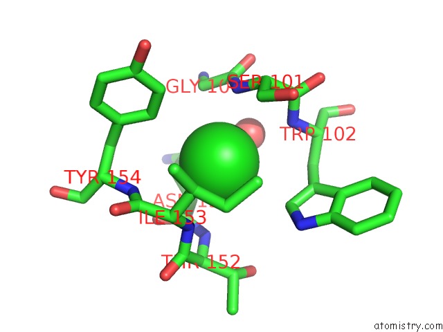
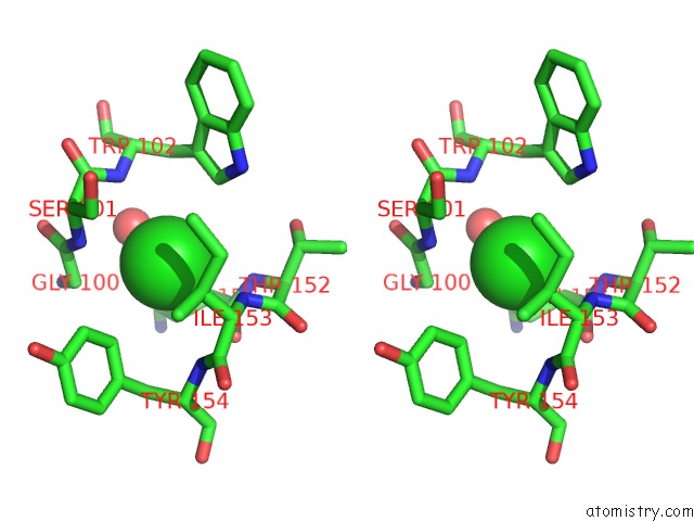
Chlorine binding site 1 out of 4 in 1w23
Go back to
Chlorine binding site 1 out
of 4 in the Crystal Structure of Phosphoserine Aminotransferase From Bacillus Alcalophilus
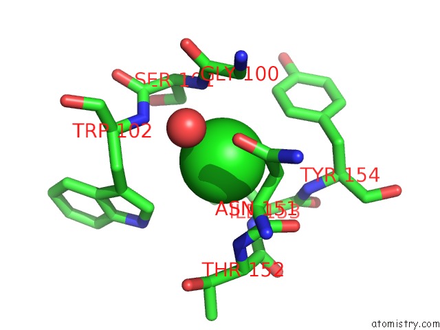
Mono view
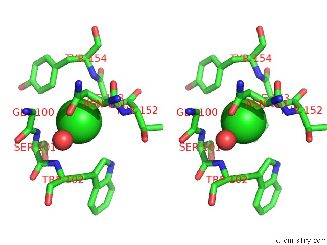
Stereo pair view
Mono view
Stereo pair view
A full contact list of Chlorine with other atoms in the Cl binding
site number 1 of Crystal Structure of Phosphoserine Aminotransferase From Bacillus Alcalophilus within 5.0Å range:
|
Chlorine binding site 2 out of 4 in 1w23
Go back to
Chlorine binding site 2 out
of 4 in the Crystal Structure of Phosphoserine Aminotransferase From Bacillus Alcalophilus
Mono view
Stereo pair view
Mono view
Stereo pair view
A full contact list of Chlorine with other atoms in the Cl binding
site number 2 of Crystal Structure of Phosphoserine Aminotransferase From Bacillus Alcalophilus within 5.0Å range:
|
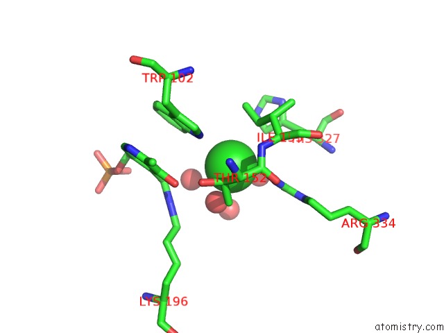
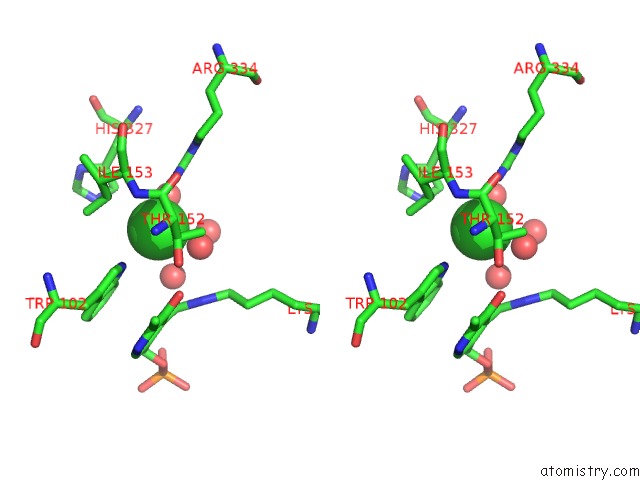
Chlorine binding site 3 out of 4 in 1w23
Go back to
Chlorine binding site 3 out
of 4 in the Crystal Structure of Phosphoserine Aminotransferase From Bacillus Alcalophilus
Mono view
Stereo pair view
Mono view
Stereo pair view
A full contact list of Chlorine with other atoms in the Cl binding
site number 3 of Crystal Structure of Phosphoserine Aminotransferase From Bacillus Alcalophilus within 5.0Å range:
|
Chlorine binding site 4 out of 4 in 1w23
Go back to
Chlorine binding site 4 out
of 4 in the Crystal Structure of Phosphoserine Aminotransferase From Bacillus Alcalophilus
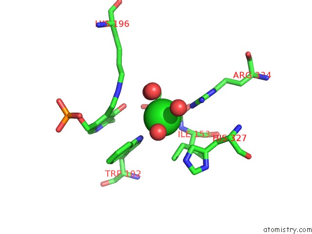
Mono view
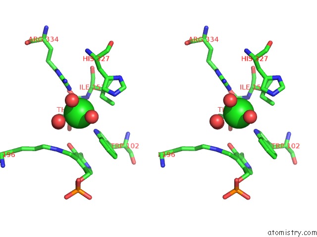
Stereo pair view
Mono view
Stereo pair view
A full contact list of Chlorine with other atoms in the Cl binding
site number 4 of Crystal Structure of Phosphoserine Aminotransferase From Bacillus Alcalophilus within 5.0Å range:
|
Reference:
A.Dubnovitsky,
E.G.Kapetaniou,
A.C.Papageorgiou.
Enzyme Adaptation to Alkaline pH: Atomic Resolution (1.08 A) Structure of Phosphoserine Aminotransferase From Bacillus Alcalophilus Protein Sci. V. 14 97 2005.
ISSN: ISSN 0961-8368
PubMed: 15608117
DOI: 10.1110/PS.041029805
Page generated: Sat Jul 20 03:30:37 2024
ISSN: ISSN 0961-8368
PubMed: 15608117
DOI: 10.1110/PS.041029805
Last articles
Zn in 9J0NZn in 9J0O
Zn in 9J0P
Zn in 9FJX
Zn in 9EKB
Zn in 9C0F
Zn in 9CAH
Zn in 9CH0
Zn in 9CH3
Zn in 9CH1