Chlorine »
PDB 1wbq-1wvf »
1wbq »
Chlorine in PDB 1wbq: Znmg Substituted Aminopeptidase P From E. Coli
Enzymatic activity of Znmg Substituted Aminopeptidase P From E. Coli
All present enzymatic activity of Znmg Substituted Aminopeptidase P From E. Coli:
3.4.11.9;
3.4.11.9;
Protein crystallography data
The structure of Znmg Substituted Aminopeptidase P From E. Coli, PDB code: 1wbq
was solved by
S.C.Graham,
C.S.Bond,
H.C.Freeman,
J.M.Guss,
with X-Ray Crystallography technique. A brief refinement statistics is given in the table below:
| Resolution Low / High (Å) | 29.97 / 2.30 |
| Space group | C 1 2 1 |
| Cell size a, b, c (Å), α, β, γ (°) | 111.974, 236.703, 137.637, 90.00, 106.14, 90.00 |
| R / Rfree (%) | 16.7 / 19.7 |
Other elements in 1wbq:
The structure of Znmg Substituted Aminopeptidase P From E. Coli also contains other interesting chemical elements:
| Magnesium | (Mg) | 6 atoms |
| Zinc | (Zn) | 4 atoms |
Chlorine Binding Sites:
The binding sites of Chlorine atom in the Znmg Substituted Aminopeptidase P From E. Coli
(pdb code 1wbq). This binding sites where shown within
5.0 Angstroms radius around Chlorine atom.
In total 4 binding sites of Chlorine where determined in the Znmg Substituted Aminopeptidase P From E. Coli, PDB code: 1wbq:
Jump to Chlorine binding site number: 1; 2; 3; 4;
In total 4 binding sites of Chlorine where determined in the Znmg Substituted Aminopeptidase P From E. Coli, PDB code: 1wbq:
Jump to Chlorine binding site number: 1; 2; 3; 4;
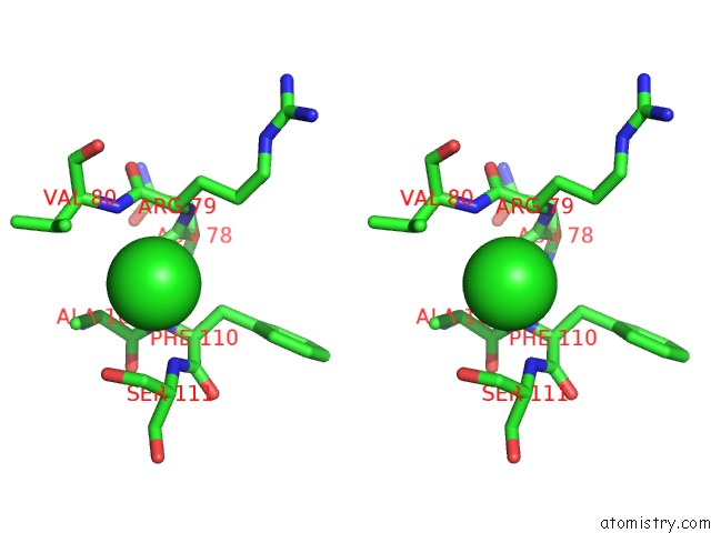
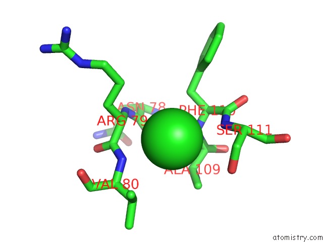
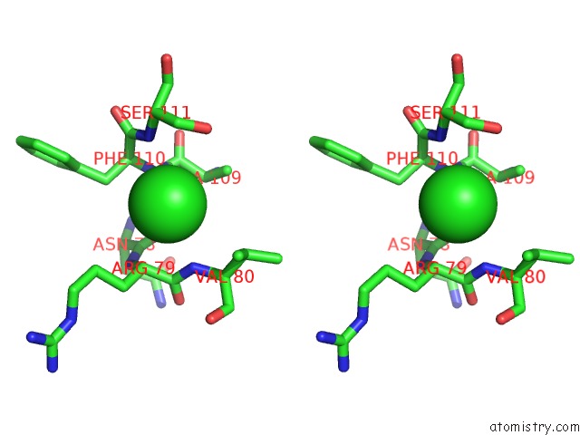
Chlorine binding site 1 out of 4 in 1wbq
Go back to
Chlorine binding site 1 out
of 4 in the Znmg Substituted Aminopeptidase P From E. Coli
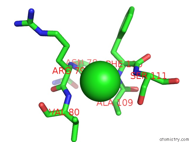
Mono view
Stereo pair view
Mono view
Stereo pair view
A full contact list of Chlorine with other atoms in the Cl binding
site number 1 of Znmg Substituted Aminopeptidase P From E. Coli within 5.0Å range:
|
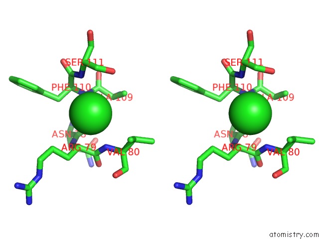
Chlorine binding site 2 out of 4 in 1wbq
Go back to
Chlorine binding site 2 out
of 4 in the Znmg Substituted Aminopeptidase P From E. Coli

Mono view
Stereo pair view
Mono view
Stereo pair view
A full contact list of Chlorine with other atoms in the Cl binding
site number 2 of Znmg Substituted Aminopeptidase P From E. Coli within 5.0Å range:
|
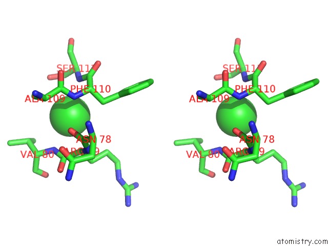
Chlorine binding site 3 out of 4 in 1wbq
Go back to
Chlorine binding site 3 out
of 4 in the Znmg Substituted Aminopeptidase P From E. Coli
Mono view
Stereo pair view
Mono view
Stereo pair view
A full contact list of Chlorine with other atoms in the Cl binding
site number 3 of Znmg Substituted Aminopeptidase P From E. Coli within 5.0Å range:
|
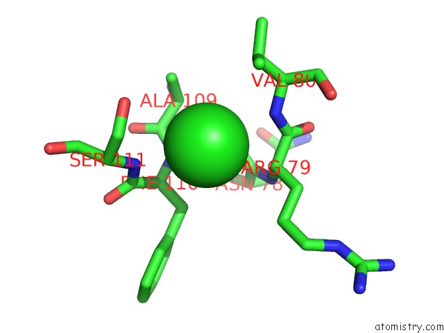
Chlorine binding site 4 out of 4 in 1wbq
Go back to
Chlorine binding site 4 out
of 4 in the Znmg Substituted Aminopeptidase P From E. Coli
Mono view
Stereo pair view
Mono view
Stereo pair view
A full contact list of Chlorine with other atoms in the Cl binding
site number 4 of Znmg Substituted Aminopeptidase P From E. Coli within 5.0Å range:
|
Reference:
S.C.Graham,
C.S.Bond,
H.C.Freeman,
J.M.Guss.
Structural and Functional Implications of Metal Ion Selection in Aminopeptidase P, A Metalloprotease with A Dinuclear Metal Center. Biochemistry V. 44 13820 2005.
ISSN: ISSN 0006-2960
PubMed: 16229471
DOI: 10.1021/BI0512849
Page generated: Thu Jul 10 20:16:19 2025
ISSN: ISSN 0006-2960
PubMed: 16229471
DOI: 10.1021/BI0512849
Last articles
Cl in 8C4PCl in 8C7M
Cl in 8C78
Cl in 8C6T
Cl in 8C61
Cl in 8C6I
Cl in 8C4Q
Cl in 8C4R
Cl in 8C5Q
Cl in 8C5N