Chlorine »
PDB 1wbq-1wvf »
1we1 »
Chlorine in PDB 1we1: Crystal Structure of Heme Oxygenase-1 From Cyanobacterium Synechocystis Sp. PCC6803 in Complex with Heme
Enzymatic activity of Crystal Structure of Heme Oxygenase-1 From Cyanobacterium Synechocystis Sp. PCC6803 in Complex with Heme
All present enzymatic activity of Crystal Structure of Heme Oxygenase-1 From Cyanobacterium Synechocystis Sp. PCC6803 in Complex with Heme:
1.14.99.3;
1.14.99.3;
Protein crystallography data
The structure of Crystal Structure of Heme Oxygenase-1 From Cyanobacterium Synechocystis Sp. PCC6803 in Complex with Heme, PDB code: 1we1
was solved by
M.Sugishima,
C.T.Migita,
X.Zhang,
T.Yoshida,
K.Fukuyama,
with X-Ray Crystallography technique. A brief refinement statistics is given in the table below:
| Resolution Low / High (Å) | 20.00 / 2.50 |
| Space group | C 1 2 1 |
| Cell size a, b, c (Å), α, β, γ (°) | 110.790, 113.730, 109.700, 90.00, 112.26, 90.00 |
| R / Rfree (%) | 22 / 26.9 |
Other elements in 1we1:
The structure of Crystal Structure of Heme Oxygenase-1 From Cyanobacterium Synechocystis Sp. PCC6803 in Complex with Heme also contains other interesting chemical elements:
| Iron | (Fe) | 4 atoms |
Chlorine Binding Sites:
The binding sites of Chlorine atom in the Crystal Structure of Heme Oxygenase-1 From Cyanobacterium Synechocystis Sp. PCC6803 in Complex with Heme
(pdb code 1we1). This binding sites where shown within
5.0 Angstroms radius around Chlorine atom.
In total 5 binding sites of Chlorine where determined in the Crystal Structure of Heme Oxygenase-1 From Cyanobacterium Synechocystis Sp. PCC6803 in Complex with Heme, PDB code: 1we1:
Jump to Chlorine binding site number: 1; 2; 3; 4; 5;
In total 5 binding sites of Chlorine where determined in the Crystal Structure of Heme Oxygenase-1 From Cyanobacterium Synechocystis Sp. PCC6803 in Complex with Heme, PDB code: 1we1:
Jump to Chlorine binding site number: 1; 2; 3; 4; 5;
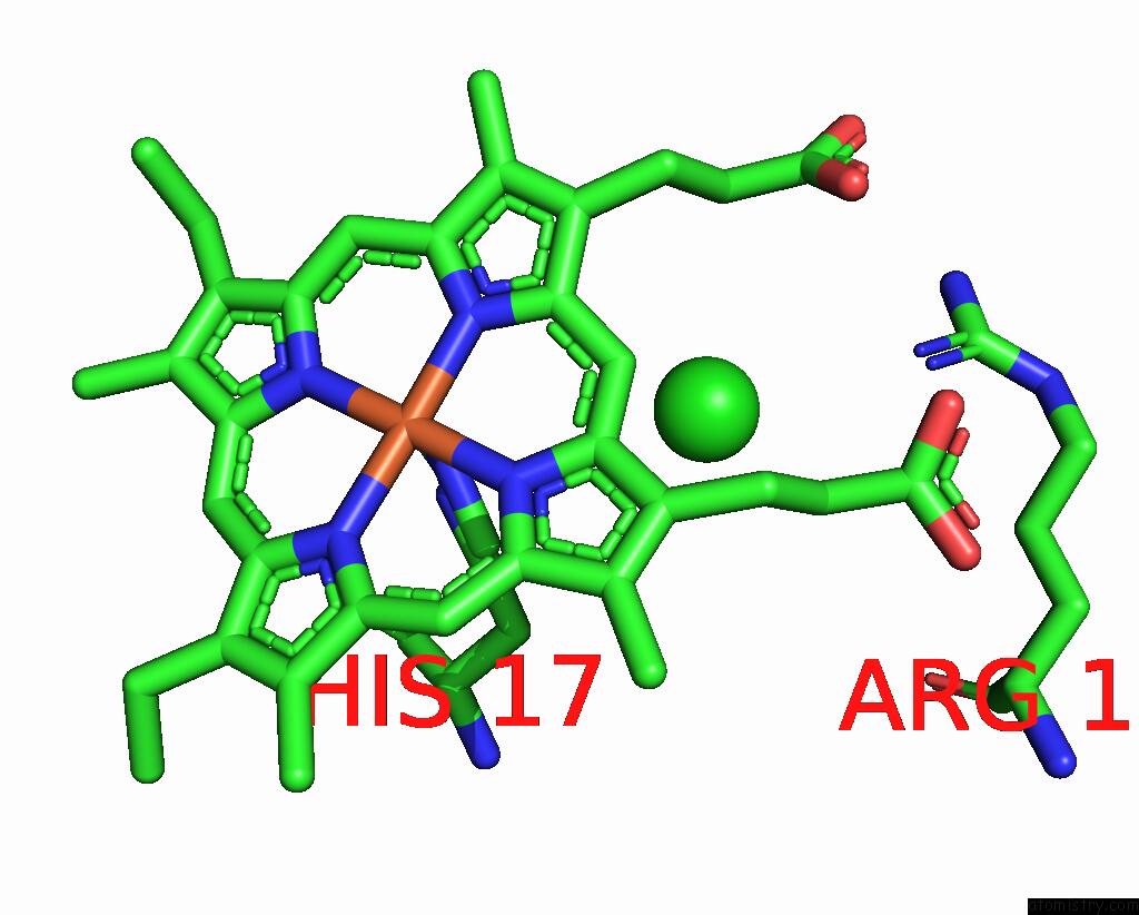
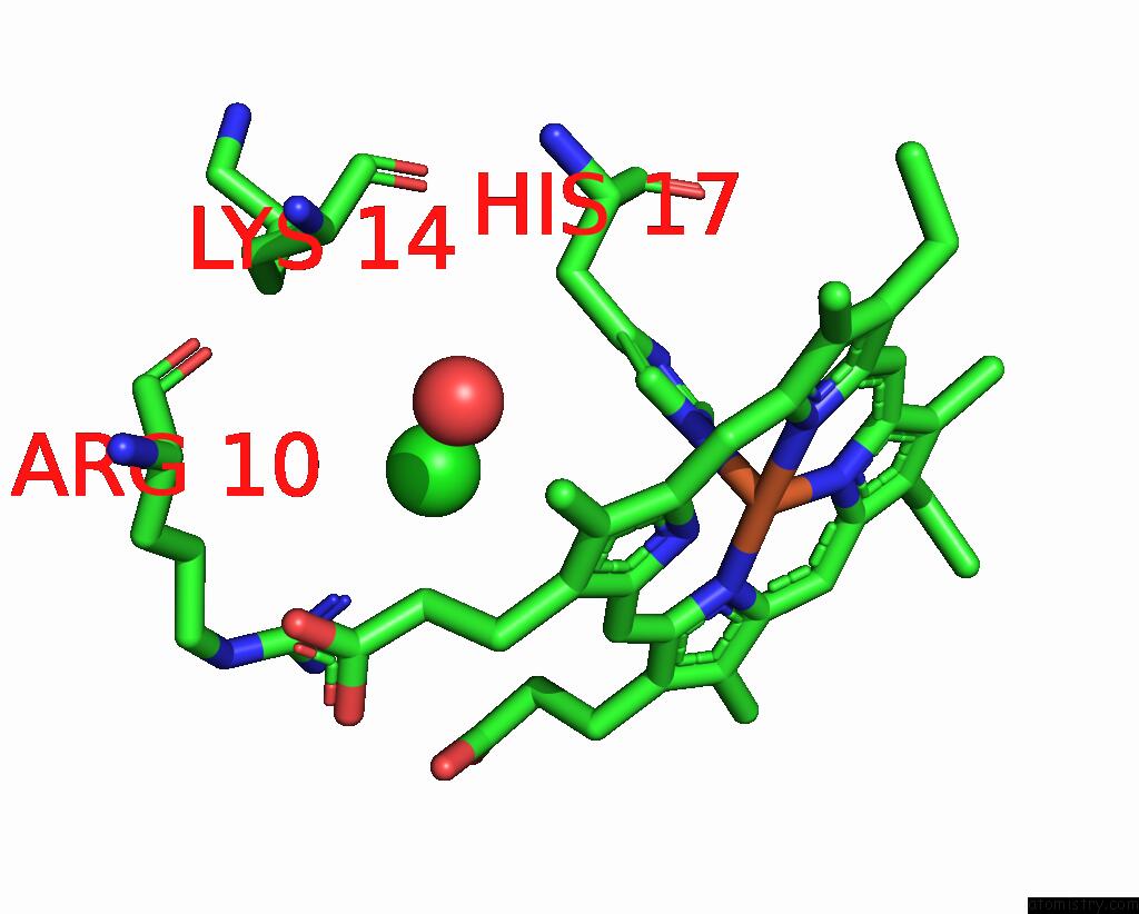
Chlorine binding site 1 out of 5 in 1we1
Go back to
Chlorine binding site 1 out
of 5 in the Crystal Structure of Heme Oxygenase-1 From Cyanobacterium Synechocystis Sp. PCC6803 in Complex with Heme
Mono view
Stereo pair view
Mono view
Stereo pair view
A full contact list of Chlorine with other atoms in the Cl binding
site number 1 of Crystal Structure of Heme Oxygenase-1 From Cyanobacterium Synechocystis Sp. PCC6803 in Complex with Heme within 5.0Å range:
|
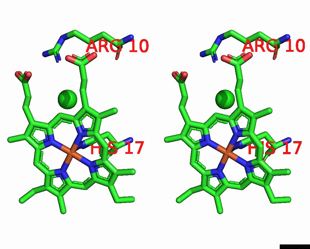
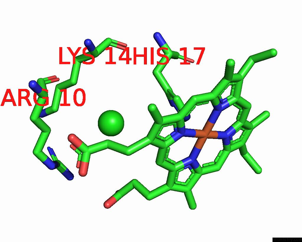
Chlorine binding site 2 out of 5 in 1we1
Go back to
Chlorine binding site 2 out
of 5 in the Crystal Structure of Heme Oxygenase-1 From Cyanobacterium Synechocystis Sp. PCC6803 in Complex with Heme
Mono view

Stereo pair view
Mono view
Stereo pair view
A full contact list of Chlorine with other atoms in the Cl binding
site number 2 of Crystal Structure of Heme Oxygenase-1 From Cyanobacterium Synechocystis Sp. PCC6803 in Complex with Heme within 5.0Å range:
|
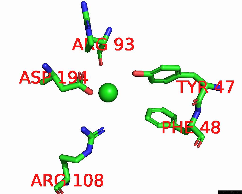
Chlorine binding site 3 out of 5 in 1we1
Go back to
Chlorine binding site 3 out
of 5 in the Crystal Structure of Heme Oxygenase-1 From Cyanobacterium Synechocystis Sp. PCC6803 in Complex with Heme

Mono view
Stereo pair view
Mono view
Stereo pair view
A full contact list of Chlorine with other atoms in the Cl binding
site number 3 of Crystal Structure of Heme Oxygenase-1 From Cyanobacterium Synechocystis Sp. PCC6803 in Complex with Heme within 5.0Å range:
|
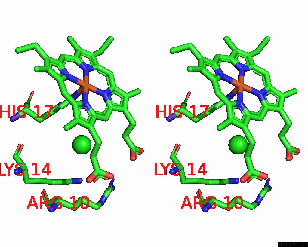
Chlorine binding site 4 out of 5 in 1we1
Go back to
Chlorine binding site 4 out
of 5 in the Crystal Structure of Heme Oxygenase-1 From Cyanobacterium Synechocystis Sp. PCC6803 in Complex with Heme
Mono view

Stereo pair view
Mono view
Stereo pair view
A full contact list of Chlorine with other atoms in the Cl binding
site number 4 of Crystal Structure of Heme Oxygenase-1 From Cyanobacterium Synechocystis Sp. PCC6803 in Complex with Heme within 5.0Å range:
|
Chlorine binding site 5 out of 5 in 1we1
Go back to
Chlorine binding site 5 out
of 5 in the Crystal Structure of Heme Oxygenase-1 From Cyanobacterium Synechocystis Sp. PCC6803 in Complex with Heme
Mono view

Stereo pair view
Mono view
Stereo pair view
A full contact list of Chlorine with other atoms in the Cl binding
site number 5 of Crystal Structure of Heme Oxygenase-1 From Cyanobacterium Synechocystis Sp. PCC6803 in Complex with Heme within 5.0Å range:
|
Reference:
M.Sugishima,
C.T.Migita,
X.Zhang,
T.Yoshida,
K.Fukuyama.
Crystal Structure of Heme Oxygenase-1 From Cyanobacterium Synechocystis Sp. Pcc 6803 in Complex with Heme Eur.J.Biochem. V. 271 4517 2004.
ISSN: ISSN 0014-2956
PubMed: 15560792
DOI: 10.1111/J.1432-1033.2004.04411.X
Page generated: Thu Jul 10 20:16:35 2025
ISSN: ISSN 0014-2956
PubMed: 15560792
DOI: 10.1111/J.1432-1033.2004.04411.X
Last articles
F in 7QN0F in 7QN4
F in 7QN1
F in 7QN2
F in 7QN3
F in 7QMY
F in 7QMX
F in 7QMW
F in 7QMZ
F in 7QMU