Chlorine »
PDB 1xkn-1y6q »
1xrl »
Chlorine in PDB 1xrl: Crystal Structure of Active Site F1-Mutant Y205F Complex with Inhibitor Pck
Enzymatic activity of Crystal Structure of Active Site F1-Mutant Y205F Complex with Inhibitor Pck
All present enzymatic activity of Crystal Structure of Active Site F1-Mutant Y205F Complex with Inhibitor Pck:
3.4.11.5;
3.4.11.5;
Protein crystallography data
The structure of Crystal Structure of Active Site F1-Mutant Y205F Complex with Inhibitor Pck, PDB code: 1xrl
was solved by
P.Goettig,
H.Brandstetter,
M.Groll,
W.Goehring,
P.V.Konarev,
D.I.Svergun,
R.Huber,
J.-S.Kim,
with X-Ray Crystallography technique. A brief refinement statistics is given in the table below:
| Resolution Low / High (Å) | 19.40 / 1.82 |
| Space group | P 21 21 21 |
| Cell size a, b, c (Å), α, β, γ (°) | 57.330, 61.850, 80.770, 90.00, 90.00, 90.00 |
| R / Rfree (%) | 24.1 / 27.9 |
Chlorine Binding Sites:
The binding sites of Chlorine atom in the Crystal Structure of Active Site F1-Mutant Y205F Complex with Inhibitor Pck
(pdb code 1xrl). This binding sites where shown within
5.0 Angstroms radius around Chlorine atom.
In total only one binding site of Chlorine was determined in the Crystal Structure of Active Site F1-Mutant Y205F Complex with Inhibitor Pck, PDB code: 1xrl:
In total only one binding site of Chlorine was determined in the Crystal Structure of Active Site F1-Mutant Y205F Complex with Inhibitor Pck, PDB code: 1xrl:
Chlorine binding site 1 out of 1 in 1xrl
Go back to
Chlorine binding site 1 out
of 1 in the Crystal Structure of Active Site F1-Mutant Y205F Complex with Inhibitor Pck
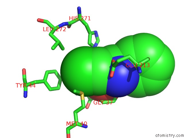
Mono view
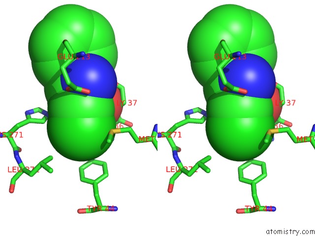
Stereo pair view
Mono view
Stereo pair view
A full contact list of Chlorine with other atoms in the Cl binding
site number 1 of Crystal Structure of Active Site F1-Mutant Y205F Complex with Inhibitor Pck within 5.0Å range:
|
Reference:
P.Goettig,
H.Brandstetter,
M.Groll,
W.Goehring,
P.V.Konarev,
D.I.Svergun,
R.Huber,
J.-S.Kim.
X-Ray Snapshots of Peptide Processing in Mutants of Tricorn-Interacting Factor F1 From Thermoplasma Acidophilum J.Biol.Chem. V. 280 33387 2005.
ISSN: ISSN 0021-9258
PubMed: 15994304
DOI: 10.1074/JBC.M505030200
Page generated: Sat Jul 20 04:03:06 2024
ISSN: ISSN 0021-9258
PubMed: 15994304
DOI: 10.1074/JBC.M505030200
Last articles
Zn in 9J0NZn in 9J0O
Zn in 9J0P
Zn in 9FJX
Zn in 9EKB
Zn in 9C0F
Zn in 9CAH
Zn in 9CH0
Zn in 9CH3
Zn in 9CH1