Chlorine »
PDB 1ymd-1zch »
1ys0 »
Chlorine in PDB 1ys0: Crystal Structure of the CDC25B Phosphatase Catalytic Domain with the Active Site Cysteine in the Disulfide Form
Enzymatic activity of Crystal Structure of the CDC25B Phosphatase Catalytic Domain with the Active Site Cysteine in the Disulfide Form
All present enzymatic activity of Crystal Structure of the CDC25B Phosphatase Catalytic Domain with the Active Site Cysteine in the Disulfide Form:
3.1.3.48;
3.1.3.48;
Protein crystallography data
The structure of Crystal Structure of the CDC25B Phosphatase Catalytic Domain with the Active Site Cysteine in the Disulfide Form, PDB code: 1ys0
was solved by
G.K.Buhrman,
B.Parker,
J.Sohn,
J.Rudolph,
C.Mattos,
with X-Ray Crystallography technique. A brief refinement statistics is given in the table below:
| Resolution Low / High (Å) | 29.70 / 2.00 |
| Space group | P 21 21 21 |
| Cell size a, b, c (Å), α, β, γ (°) | 50.197, 71.156, 73.693, 90.00, 90.00, 90.00 |
| R / Rfree (%) | 20.9 / 24.4 |
Chlorine Binding Sites:
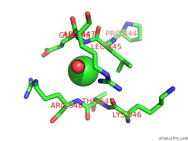
The binding sites of Chlorine atom in the Crystal Structure of the CDC25B Phosphatase Catalytic Domain with the Active Site Cysteine in the Disulfide Form
(pdb code 1ys0). This binding sites where shown within
5.0 Angstroms radius around Chlorine atom.
In total only one binding site of Chlorine was determined in the Crystal Structure of the CDC25B Phosphatase Catalytic Domain with the Active Site Cysteine in the Disulfide Form, PDB code: 1ys0:
In total only one binding site of Chlorine was determined in the Crystal Structure of the CDC25B Phosphatase Catalytic Domain with the Active Site Cysteine in the Disulfide Form, PDB code: 1ys0:
Chlorine binding site 1 out of 1 in 1ys0
Go back to
Chlorine binding site 1 out
of 1 in the Crystal Structure of the CDC25B Phosphatase Catalytic Domain with the Active Site Cysteine in the Disulfide Form
Mono view
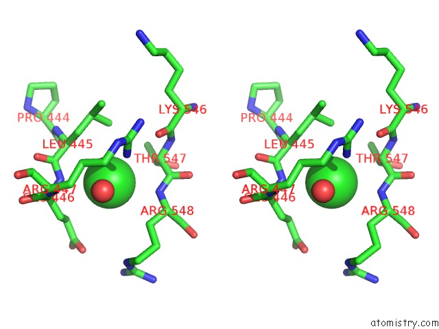
Stereo pair view
Mono view
Stereo pair view
A full contact list of Chlorine with other atoms in the Cl binding
site number 1 of Crystal Structure of the CDC25B Phosphatase Catalytic Domain with the Active Site Cysteine in the Disulfide Form within 5.0Å range:
|
Reference:
G.K.Buhrman,
B.Parker,
J.Sohn,
J.Rudolph,
C.Mattos.
Structural Mechanism of Oxidative Regulation of the Phosphatase CDC25B Via An Intramolecular Disulfide Bond Biochemistry V. 44 5307 2005.
ISSN: ISSN 0006-2960
PubMed: 15807524
DOI: 10.1021/BI047449FS0006-2960(04)07449-5
Page generated: Sat Jul 20 04:30:37 2024
ISSN: ISSN 0006-2960
PubMed: 15807524
DOI: 10.1021/BI047449FS0006-2960(04)07449-5
Last articles
Zn in 9J0NZn in 9J0O
Zn in 9J0P
Zn in 9FJX
Zn in 9EKB
Zn in 9C0F
Zn in 9CAH
Zn in 9CH0
Zn in 9CH3
Zn in 9CH1