Chlorine »
PDB 1zop-232l »
1zov »
Chlorine in PDB 1zov: Crystal Structure of Monomeric Sarcosine Oxidase From Bacillus Sp. Ns- 129
Enzymatic activity of Crystal Structure of Monomeric Sarcosine Oxidase From Bacillus Sp. Ns- 129
All present enzymatic activity of Crystal Structure of Monomeric Sarcosine Oxidase From Bacillus Sp. Ns- 129:
1.5.3.1;
1.5.3.1;
Protein crystallography data
The structure of Crystal Structure of Monomeric Sarcosine Oxidase From Bacillus Sp. Ns- 129, PDB code: 1zov
was solved by
K.Nagata,
H.Sasaki,
J.Ohtsuka,
M.Hua,
M.Okai,
K.Kubota,
M.Kamo,
K.Ito,
T.Ichikawa,
Y.Koyama,
M.Tanokura,
with X-Ray Crystallography technique. A brief refinement statistics is given in the table below:
| Resolution Low / High (Å) | 8.00 / 1.86 |
| Space group | P 41 21 2 |
| Cell size a, b, c (Å), α, β, γ (°) | 171.042, 171.042, 72.455, 90.00, 90.00, 90.00 |
| R / Rfree (%) | 15.8 / 19.1 |
Chlorine Binding Sites:
The binding sites of Chlorine atom in the Crystal Structure of Monomeric Sarcosine Oxidase From Bacillus Sp. Ns- 129
(pdb code 1zov). This binding sites where shown within
5.0 Angstroms radius around Chlorine atom.
In total 2 binding sites of Chlorine where determined in the Crystal Structure of Monomeric Sarcosine Oxidase From Bacillus Sp. Ns- 129, PDB code: 1zov:
Jump to Chlorine binding site number: 1; 2;
In total 2 binding sites of Chlorine where determined in the Crystal Structure of Monomeric Sarcosine Oxidase From Bacillus Sp. Ns- 129, PDB code: 1zov:
Jump to Chlorine binding site number: 1; 2;
Chlorine binding site 1 out of 2 in 1zov
Go back to
Chlorine binding site 1 out
of 2 in the Crystal Structure of Monomeric Sarcosine Oxidase From Bacillus Sp. Ns- 129
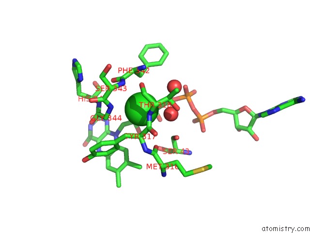
Mono view
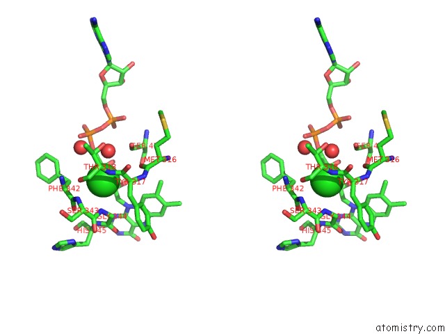
Stereo pair view
Mono view
Stereo pair view
A full contact list of Chlorine with other atoms in the Cl binding
site number 1 of Crystal Structure of Monomeric Sarcosine Oxidase From Bacillus Sp. Ns- 129 within 5.0Å range:
|
Chlorine binding site 2 out of 2 in 1zov
Go back to
Chlorine binding site 2 out
of 2 in the Crystal Structure of Monomeric Sarcosine Oxidase From Bacillus Sp. Ns- 129
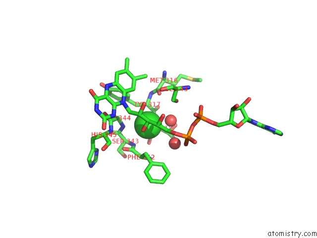
Mono view
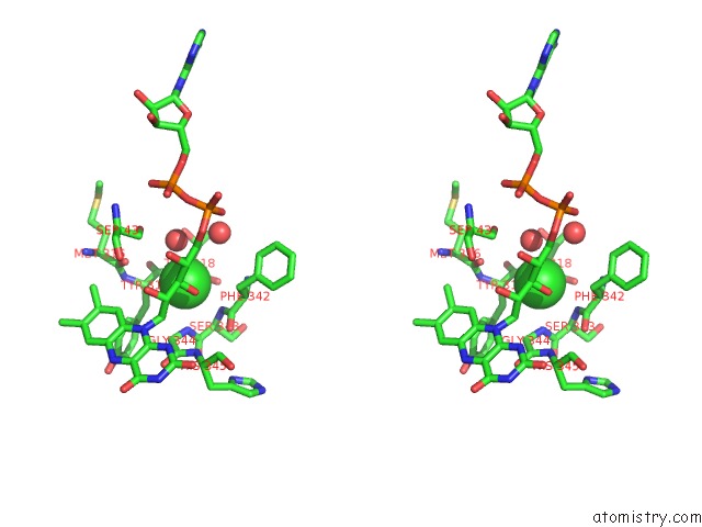
Stereo pair view
Mono view
Stereo pair view
A full contact list of Chlorine with other atoms in the Cl binding
site number 2 of Crystal Structure of Monomeric Sarcosine Oxidase From Bacillus Sp. Ns- 129 within 5.0Å range:
|
Reference:
K.Nagata,
H.Sasaki,
M.Hua,
M.Okai,
K.Kubota,
M.Kamo,
K.Ito,
T.Ichikawa,
Y.Koyama,
M.Tanokura.
Crystal Structure of Monomeric Sarcosine Oxidase From Bacillus Sp. Ns-129 Reveals Multiple Conformations at the Active-Site Loop Proc.Jpn.Acad.,Ser.B V. 81 220 2005.
ISSN: ISSN 0386-2208
Page generated: Sat Jul 20 04:48:24 2024
ISSN: ISSN 0386-2208
Last articles
Zn in 9J0NZn in 9J0O
Zn in 9J0P
Zn in 9FJX
Zn in 9EKB
Zn in 9C0F
Zn in 9CAH
Zn in 9CH0
Zn in 9CH3
Zn in 9CH1