Chlorine »
PDB 2a7d-2aj4 »
2a9b »
Chlorine in PDB 2a9b: Crystal Structure of R138Q Mutant of Recombinant Sulfite Oxidase at Resting State
Enzymatic activity of Crystal Structure of R138Q Mutant of Recombinant Sulfite Oxidase at Resting State
All present enzymatic activity of Crystal Structure of R138Q Mutant of Recombinant Sulfite Oxidase at Resting State:
1.8.3.1;
1.8.3.1;
Protein crystallography data
The structure of Crystal Structure of R138Q Mutant of Recombinant Sulfite Oxidase at Resting State, PDB code: 2a9b
was solved by
E.Karakas,
H.L.Wilson,
T.N.Graf,
S.Xiang,
S.Jaramillo-Busquets,
K.V.Rajagopalan,
C.Kisker,
with X-Ray Crystallography technique. A brief refinement statistics is given in the table below:
| Resolution Low / High (Å) | 30.00 / 2.50 |
| Space group | I 41 |
| Cell size a, b, c (Å), α, β, γ (°) | 86.109, 86.109, 153.825, 90.00, 90.00, 90.00 |
| R / Rfree (%) | 15.6 / 20.2 |
Other elements in 2a9b:
The structure of Crystal Structure of R138Q Mutant of Recombinant Sulfite Oxidase at Resting State also contains other interesting chemical elements:
| Molybdenum | (Mo) | 1 atom |
Chlorine Binding Sites:
The binding sites of Chlorine atom in the Crystal Structure of R138Q Mutant of Recombinant Sulfite Oxidase at Resting State
(pdb code 2a9b). This binding sites where shown within
5.0 Angstroms radius around Chlorine atom.
In total only one binding site of Chlorine was determined in the Crystal Structure of R138Q Mutant of Recombinant Sulfite Oxidase at Resting State, PDB code: 2a9b:
In total only one binding site of Chlorine was determined in the Crystal Structure of R138Q Mutant of Recombinant Sulfite Oxidase at Resting State, PDB code: 2a9b:
Chlorine binding site 1 out of 1 in 2a9b
Go back to
Chlorine binding site 1 out
of 1 in the Crystal Structure of R138Q Mutant of Recombinant Sulfite Oxidase at Resting State
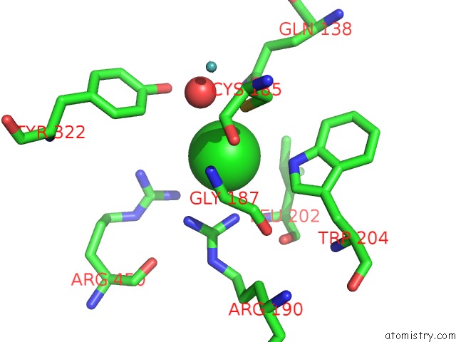
Mono view
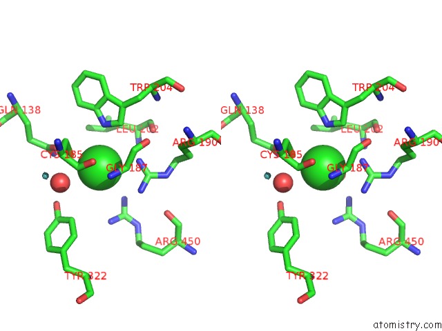
Stereo pair view
Mono view
Stereo pair view
A full contact list of Chlorine with other atoms in the Cl binding
site number 1 of Crystal Structure of R138Q Mutant of Recombinant Sulfite Oxidase at Resting State within 5.0Å range:
|
Reference:
E.Karakas,
H.L.Wilson,
T.N.Graf,
S.Xiang,
S.Jaramillo-Busquets,
K.V.Rajagopalan,
C.Kisker.
Structural Insights Into Sulfite Oxidase Deficiency J.Biol.Chem. V. 280 33506 2005.
ISSN: ISSN 0021-9258
PubMed: 16048997
DOI: 10.1074/JBC.M505035200
Page generated: Sat Jul 20 05:09:49 2024
ISSN: ISSN 0021-9258
PubMed: 16048997
DOI: 10.1074/JBC.M505035200
Last articles
Zn in 9J0NZn in 9J0O
Zn in 9J0P
Zn in 9FJX
Zn in 9EKB
Zn in 9C0F
Zn in 9CAH
Zn in 9CH0
Zn in 9CH3
Zn in 9CH1