Chlorine in PDB 2aih: 1H-uc(Nmr) Solution Structure of A Trypsin/Chymotrypsin Bowman- Birk Inhibitor From Lens Culinaris.
Chlorine Binding Sites:
Pages:
>>> Page 1 <<<
Page 2, Binding sites: 11 -
11;
Binding sites:
The binding sites of Chlorine atom in the 1H-uc(Nmr) Solution Structure of A Trypsin/Chymotrypsin Bowman- Birk Inhibitor From Lens Culinaris.
(pdb code 2aih). This binding sites where shown within
5.0 Angstroms radius around Chlorine atom.
In total 11 binding sites of Chlorine where determined in the
1H-uc(Nmr) Solution Structure of A Trypsin/Chymotrypsin Bowman- Birk Inhibitor From Lens Culinaris., PDB code: 2aih:
Jump to Chlorine binding site number:
1;
2;
3;
4;
5;
6;
7;
8;
9;
10;
Chlorine binding site 1 out
of 11 in 2aih
Go back to
Chlorine Binding Sites List in 2aih
Chlorine binding site 1 out
of 11 in the 1H-uc(Nmr) Solution Structure of A Trypsin/Chymotrypsin Bowman- Birk Inhibitor From Lens Culinaris.
Mono view
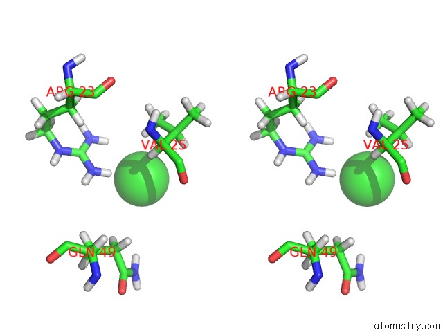
Stereo pair view
|
A full contact list of Chlorine with other atoms in the Cl binding
site number 1 of 1H-uc(Nmr) Solution Structure of A Trypsin/Chymotrypsin Bowman- Birk Inhibitor From Lens Culinaris. within 5.0Å range:
probe
|
atom
|
residue
|
distance (Å)
|
B
|
Occ
|
A:Cl68
b:0.0
occ:1.00
|
HH22
|
A:ARG23
|
2.9
|
0.0
|
1.0
|
HH12
|
A:ARG23
|
3.4
|
0.0
|
1.0
|
HG13
|
A:VAL25
|
3.5
|
0.0
|
1.0
|
NH2
|
A:ARG23
|
3.9
|
0.0
|
1.0
|
HA
|
A:VAL25
|
4.2
|
0.0
|
1.0
|
NH1
|
A:ARG23
|
4.2
|
0.0
|
1.0
|
HB
|
A:VAL25
|
4.3
|
0.0
|
1.0
|
CG1
|
A:VAL25
|
4.4
|
0.0
|
1.0
|
CZ
|
A:ARG23
|
4.5
|
0.0
|
1.0
|
HH21
|
A:ARG23
|
4.6
|
0.0
|
1.0
|
HG11
|
A:VAL25
|
4.6
|
0.0
|
1.0
|
HG2
|
A:GLN49
|
4.7
|
0.0
|
1.0
|
CB
|
A:VAL25
|
4.8
|
0.0
|
1.0
|
|
Chlorine binding site 2 out
of 11 in 2aih
Go back to
Chlorine Binding Sites List in 2aih
Chlorine binding site 2 out
of 11 in the 1H-uc(Nmr) Solution Structure of A Trypsin/Chymotrypsin Bowman- Birk Inhibitor From Lens Culinaris.
Mono view
Stereo pair view
|
A full contact list of Chlorine with other atoms in the Cl binding
site number 2 of 1H-uc(Nmr) Solution Structure of A Trypsin/Chymotrypsin Bowman- Birk Inhibitor From Lens Culinaris. within 5.0Å range:
probe
|
atom
|
residue
|
distance (Å)
|
B
|
Occ
|
A:Cl69
b:0.0
occ:1.00
|
HD2
|
A:HIS32
|
2.7
|
0.0
|
1.0
|
H
|
A:SER33
|
3.0
|
0.0
|
1.0
|
HA
|
A:HIS32
|
3.2
|
0.0
|
1.0
|
HB3
|
A:HIS32
|
3.2
|
0.0
|
1.0
|
CD2
|
A:HIS32
|
3.6
|
0.0
|
1.0
|
CB
|
A:HIS32
|
3.8
|
0.0
|
1.0
|
CA
|
A:HIS32
|
3.9
|
0.0
|
1.0
|
N
|
A:SER33
|
4.0
|
0.0
|
1.0
|
CG
|
A:HIS32
|
4.0
|
0.0
|
1.0
|
C
|
A:HIS32
|
4.5
|
0.0
|
1.0
|
O
|
A:SER6
|
4.5
|
0.0
|
1.0
|
HB2
|
A:SER33
|
4.6
|
0.0
|
1.0
|
HB3
|
A:SER33
|
4.7
|
0.0
|
1.0
|
NE2
|
A:HIS32
|
4.8
|
0.0
|
1.0
|
HB2
|
A:HIS32
|
4.9
|
0.0
|
1.0
|
HA
|
A:SER6
|
4.9
|
0.0
|
1.0
|
H
|
A:ALA34
|
5.0
|
0.0
|
1.0
|
|
Chlorine binding site 3 out
of 11 in 2aih
Go back to
Chlorine Binding Sites List in 2aih
Chlorine binding site 3 out
of 11 in the 1H-uc(Nmr) Solution Structure of A Trypsin/Chymotrypsin Bowman- Birk Inhibitor From Lens Culinaris.
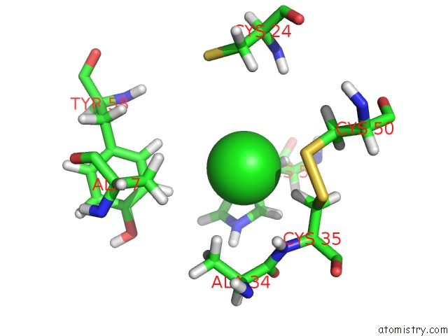
Mono view
Stereo pair view
|
A full contact list of Chlorine with other atoms in the Cl binding
site number 3 of 1H-uc(Nmr) Solution Structure of A Trypsin/Chymotrypsin Bowman- Birk Inhibitor From Lens Culinaris. within 5.0Å range:
probe
|
atom
|
residue
|
distance (Å)
|
B
|
Occ
|
A:Cl70
b:0.0
occ:1.00
|
HB3
|
A:CYS35
|
2.9
|
0.0
|
1.0
|
CD2
|
A:HIS54
|
3.3
|
0.0
|
1.0
|
HD2
|
A:HIS54
|
3.3
|
0.0
|
1.0
|
HB2
|
A:HIS54
|
3.4
|
0.0
|
1.0
|
CG
|
A:HIS54
|
3.5
|
0.0
|
1.0
|
NE2
|
A:HIS54
|
3.7
|
0.0
|
1.0
|
HE1
|
A:TYR58
|
3.9
|
0.0
|
1.0
|
CB
|
A:HIS54
|
3.9
|
0.0
|
1.0
|
CB
|
A:CYS35
|
4.0
|
0.0
|
1.0
|
HB2
|
A:ALA34
|
4.0
|
0.0
|
1.0
|
ND1
|
A:HIS54
|
4.1
|
0.0
|
1.0
|
HE2
|
A:HIS54
|
4.1
|
0.0
|
1.0
|
HB3
|
A:HIS54
|
4.1
|
0.0
|
1.0
|
CE1
|
A:HIS54
|
4.2
|
0.0
|
1.0
|
SG
|
A:CYS50
|
4.3
|
0.0
|
1.0
|
HD1
|
A:TYR58
|
4.3
|
0.0
|
1.0
|
N
|
A:CYS35
|
4.4
|
0.0
|
1.0
|
H
|
A:CYS35
|
4.4
|
0.0
|
1.0
|
HB1
|
A:ALA34
|
4.4
|
0.0
|
1.0
|
HB2
|
A:CYS35
|
4.5
|
0.0
|
1.0
|
HD1
|
A:HIS54
|
4.6
|
0.0
|
1.0
|
HA
|
A:CYS35
|
4.6
|
0.0
|
1.0
|
CA
|
A:CYS35
|
4.6
|
0.0
|
1.0
|
HB3
|
A:CYS50
|
4.7
|
0.0
|
1.0
|
CB
|
A:ALA34
|
4.7
|
0.0
|
1.0
|
C
|
A:ALA34
|
4.8
|
0.0
|
1.0
|
CE1
|
A:TYR58
|
4.8
|
0.0
|
1.0
|
HE1
|
A:HIS54
|
4.9
|
0.0
|
1.0
|
SG
|
A:CYS35
|
4.9
|
0.0
|
1.0
|
HB2
|
A:CYS24
|
4.9
|
0.0
|
1.0
|
HB2
|
A:ALA7
|
4.9
|
0.0
|
1.0
|
CD1
|
A:TYR58
|
5.0
|
0.0
|
1.0
|
HB2
|
A:CYS50
|
5.0
|
0.0
|
1.0
|
CB
|
A:CYS50
|
5.0
|
0.0
|
1.0
|
HB1
|
A:ALA7
|
5.0
|
0.0
|
1.0
|
|
Chlorine binding site 4 out
of 11 in 2aih
Go back to
Chlorine Binding Sites List in 2aih
Chlorine binding site 4 out
of 11 in the 1H-uc(Nmr) Solution Structure of A Trypsin/Chymotrypsin Bowman- Birk Inhibitor From Lens Culinaris.
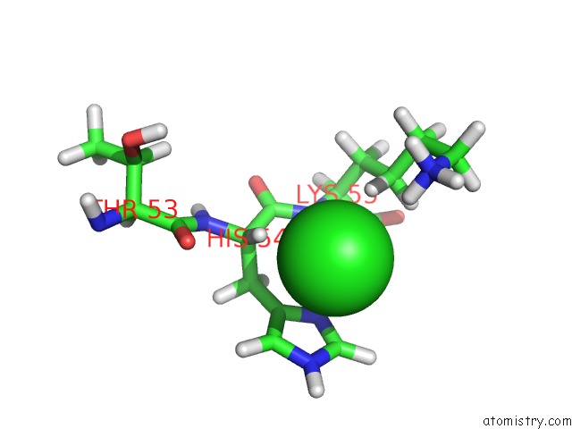
Mono view
Stereo pair view
|
A full contact list of Chlorine with other atoms in the Cl binding
site number 4 of 1H-uc(Nmr) Solution Structure of A Trypsin/Chymotrypsin Bowman- Birk Inhibitor From Lens Culinaris. within 5.0Å range:
probe
|
atom
|
residue
|
distance (Å)
|
B
|
Occ
|
A:Cl71
b:0.0
occ:1.00
|
HZ2
|
A:LYS55
|
2.9
|
0.0
|
1.0
|
HA
|
A:HIS54
|
3.0
|
0.0
|
1.0
|
HG2
|
A:LYS55
|
3.1
|
0.0
|
1.0
|
H
|
A:LYS55
|
3.2
|
0.0
|
1.0
|
CG
|
A:HIS54
|
3.6
|
0.0
|
1.0
|
CD2
|
A:HIS54
|
3.6
|
0.0
|
1.0
|
ND1
|
A:HIS54
|
3.6
|
0.0
|
1.0
|
NE2
|
A:HIS54
|
3.7
|
0.0
|
1.0
|
CE1
|
A:HIS54
|
3.7
|
0.0
|
1.0
|
NZ
|
A:LYS55
|
3.9
|
0.0
|
1.0
|
CA
|
A:HIS54
|
3.9
|
0.0
|
1.0
|
N
|
A:LYS55
|
4.0
|
0.0
|
1.0
|
HD2
|
A:HIS54
|
4.1
|
0.0
|
1.0
|
HZ3
|
A:LYS55
|
4.1
|
0.0
|
1.0
|
HE2
|
A:HIS54
|
4.1
|
0.0
|
1.0
|
HD1
|
A:HIS54
|
4.1
|
0.0
|
1.0
|
HE1
|
A:HIS54
|
4.2
|
0.0
|
1.0
|
CG
|
A:LYS55
|
4.2
|
0.0
|
1.0
|
HZ1
|
A:LYS55
|
4.3
|
0.0
|
1.0
|
CB
|
A:HIS54
|
4.3
|
0.0
|
1.0
|
HG3
|
A:LYS55
|
4.4
|
0.0
|
1.0
|
C
|
A:HIS54
|
4.4
|
0.0
|
1.0
|
O
|
A:THR53
|
4.6
|
0.0
|
1.0
|
CE
|
A:LYS55
|
4.9
|
0.0
|
1.0
|
|
Chlorine binding site 5 out
of 11 in 2aih
Go back to
Chlorine Binding Sites List in 2aih
Chlorine binding site 5 out
of 11 in the 1H-uc(Nmr) Solution Structure of A Trypsin/Chymotrypsin Bowman- Birk Inhibitor From Lens Culinaris.
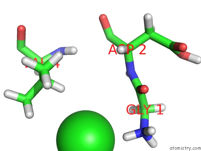
Mono view
Stereo pair view
|
A full contact list of Chlorine with other atoms in the Cl binding
site number 5 of 1H-uc(Nmr) Solution Structure of A Trypsin/Chymotrypsin Bowman- Birk Inhibitor From Lens Culinaris. within 5.0Å range:
probe
|
atom
|
residue
|
distance (Å)
|
B
|
Occ
|
A:Cl72
b:0.0
occ:1.00
|
H3
|
A:GLY1
|
2.5
|
0.0
|
1.0
|
HB
|
A:VAL4
|
3.4
|
0.0
|
1.0
|
HA3
|
A:GLY1
|
3.5
|
0.0
|
1.0
|
N
|
A:GLY1
|
3.5
|
0.0
|
1.0
|
HG22
|
A:VAL4
|
3.7
|
0.0
|
1.0
|
CA
|
A:GLY1
|
4.0
|
0.0
|
1.0
|
H2
|
A:GLY1
|
4.0
|
0.0
|
1.0
|
HG23
|
A:VAL4
|
4.1
|
0.0
|
1.0
|
H1
|
A:GLY1
|
4.1
|
0.0
|
1.0
|
CG2
|
A:VAL4
|
4.2
|
0.0
|
1.0
|
CB
|
A:VAL4
|
4.3
|
0.0
|
1.0
|
C
|
A:GLY1
|
4.4
|
0.0
|
1.0
|
HG11
|
A:VAL4
|
4.7
|
0.0
|
1.0
|
O
|
A:GLY1
|
4.9
|
0.0
|
1.0
|
N
|
A:ASP2
|
4.9
|
0.0
|
1.0
|
H
|
A:ASP2
|
4.9
|
0.0
|
1.0
|
HA2
|
A:GLY1
|
5.0
|
0.0
|
1.0
|
|
Chlorine binding site 6 out
of 11 in 2aih
Go back to
Chlorine Binding Sites List in 2aih
Chlorine binding site 6 out
of 11 in the 1H-uc(Nmr) Solution Structure of A Trypsin/Chymotrypsin Bowman- Birk Inhibitor From Lens Culinaris.
Mono view
Stereo pair view
|
A full contact list of Chlorine with other atoms in the Cl binding
site number 6 of 1H-uc(Nmr) Solution Structure of A Trypsin/Chymotrypsin Bowman- Birk Inhibitor From Lens Culinaris. within 5.0Å range:
probe
|
atom
|
residue
|
distance (Å)
|
B
|
Occ
|
A:Cl73
b:0.0
occ:1.00
|
HH
|
A:TYR42
|
2.2
|
0.0
|
1.0
|
HE1
|
A:TYR42
|
3.2
|
0.0
|
1.0
|
OH
|
A:TYR42
|
3.2
|
0.0
|
1.0
|
CE1
|
A:TYR42
|
3.9
|
0.0
|
1.0
|
CZ
|
A:TYR42
|
4.0
|
0.0
|
1.0
|
|
Chlorine binding site 7 out
of 11 in 2aih
Go back to
Chlorine Binding Sites List in 2aih
Chlorine binding site 7 out
of 11 in the 1H-uc(Nmr) Solution Structure of A Trypsin/Chymotrypsin Bowman- Birk Inhibitor From Lens Culinaris.
Mono view
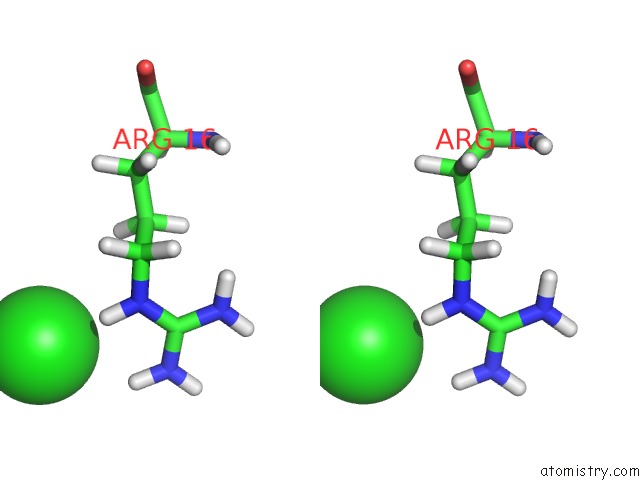
Stereo pair view
|
A full contact list of Chlorine with other atoms in the Cl binding
site number 7 of 1H-uc(Nmr) Solution Structure of A Trypsin/Chymotrypsin Bowman- Birk Inhibitor From Lens Culinaris. within 5.0Å range:
probe
|
atom
|
residue
|
distance (Å)
|
B
|
Occ
|
A:Cl74
b:0.0
occ:1.00
|
HE
|
A:ARG16
|
2.6
|
0.0
|
1.0
|
HH21
|
A:ARG16
|
3.1
|
0.0
|
1.0
|
NE
|
A:ARG16
|
3.5
|
0.0
|
1.0
|
NH2
|
A:ARG16
|
3.9
|
0.0
|
1.0
|
HG2
|
A:ARG16
|
4.1
|
0.0
|
1.0
|
CZ
|
A:ARG16
|
4.1
|
0.0
|
1.0
|
HD3
|
A:ARG16
|
4.3
|
0.0
|
1.0
|
CD
|
A:ARG16
|
4.4
|
0.0
|
1.0
|
HH22
|
A:ARG16
|
4.7
|
0.0
|
1.0
|
CG
|
A:ARG16
|
4.9
|
0.0
|
1.0
|
|
Chlorine binding site 8 out
of 11 in 2aih
Go back to
Chlorine Binding Sites List in 2aih
Chlorine binding site 8 out
of 11 in the 1H-uc(Nmr) Solution Structure of A Trypsin/Chymotrypsin Bowman- Birk Inhibitor From Lens Culinaris.
Mono view
Stereo pair view
|
A full contact list of Chlorine with other atoms in the Cl binding
site number 8 of 1H-uc(Nmr) Solution Structure of A Trypsin/Chymotrypsin Bowman- Birk Inhibitor From Lens Culinaris. within 5.0Å range:
probe
|
atom
|
residue
|
distance (Å)
|
B
|
Occ
|
A:Cl75
b:0.0
occ:1.00
|
H
|
A:HIS62
|
2.4
|
0.0
|
1.0
|
HD2
|
A:HIS62
|
2.7
|
0.0
|
1.0
|
HA
|
A:CYS61
|
3.3
|
0.0
|
1.0
|
N
|
A:HIS62
|
3.3
|
0.0
|
1.0
|
CD2
|
A:HIS62
|
3.3
|
0.0
|
1.0
|
HB2
|
A:HIS62
|
3.4
|
0.0
|
1.0
|
CG
|
A:HIS62
|
3.8
|
0.0
|
1.0
|
CB
|
A:HIS62
|
3.9
|
0.0
|
1.0
|
CA
|
A:CYS61
|
4.1
|
0.0
|
1.0
|
C
|
A:CYS61
|
4.2
|
0.0
|
1.0
|
CA
|
A:HIS62
|
4.2
|
0.0
|
1.0
|
NE2
|
A:HIS62
|
4.3
|
0.0
|
1.0
|
HB3
|
A:CYS61
|
4.4
|
0.0
|
1.0
|
HE2
|
A:HIS62
|
4.6
|
0.0
|
1.0
|
O
|
A:HIS62
|
4.7
|
0.0
|
1.0
|
CB
|
A:CYS61
|
4.9
|
0.0
|
1.0
|
ND1
|
A:HIS62
|
4.9
|
0.0
|
1.0
|
C
|
A:HIS62
|
4.9
|
0.0
|
1.0
|
HB3
|
A:HIS62
|
5.0
|
0.0
|
1.0
|
|
Chlorine binding site 9 out
of 11 in 2aih
Go back to
Chlorine Binding Sites List in 2aih
Chlorine binding site 9 out
of 11 in the 1H-uc(Nmr) Solution Structure of A Trypsin/Chymotrypsin Bowman- Birk Inhibitor From Lens Culinaris.
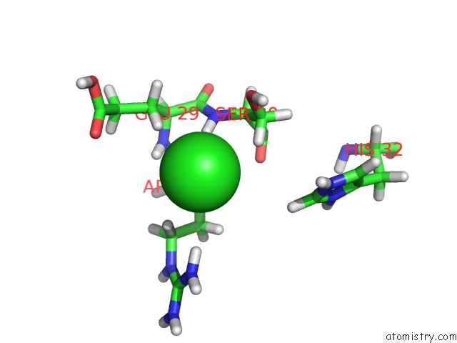
Mono view
Stereo pair view
|
A full contact list of Chlorine with other atoms in the Cl binding
site number 9 of 1H-uc(Nmr) Solution Structure of A Trypsin/Chymotrypsin Bowman- Birk Inhibitor From Lens Culinaris. within 5.0Å range:
probe
|
atom
|
residue
|
distance (Å)
|
B
|
Occ
|
A:Cl76
b:0.0
occ:1.00
|
H
|
A:SER30
|
2.7
|
0.0
|
1.0
|
HB2
|
A:SER30
|
3.0
|
0.0
|
1.0
|
HB3
|
A:ARG28
|
3.1
|
0.0
|
1.0
|
HB2
|
A:GLU29
|
3.2
|
0.0
|
1.0
|
HD2
|
A:ARG28
|
3.4
|
0.0
|
1.0
|
HH11
|
A:ARG28
|
3.5
|
0.0
|
1.0
|
N
|
A:SER30
|
3.7
|
0.0
|
1.0
|
CB
|
A:SER30
|
3.8
|
0.0
|
1.0
|
H
|
A:GLU29
|
3.9
|
0.0
|
1.0
|
OG
|
A:SER30
|
4.0
|
0.0
|
1.0
|
HG3
|
A:ARG28
|
4.0
|
0.0
|
1.0
|
HG
|
A:SER30
|
4.1
|
0.0
|
1.0
|
CB
|
A:ARG28
|
4.1
|
0.0
|
1.0
|
CA
|
A:SER30
|
4.2
|
0.0
|
1.0
|
CB
|
A:GLU29
|
4.3
|
0.0
|
1.0
|
CD
|
A:ARG28
|
4.3
|
0.0
|
1.0
|
CG
|
A:ARG28
|
4.4
|
0.0
|
1.0
|
HE1
|
A:HIS32
|
4.5
|
0.0
|
1.0
|
NH1
|
A:ARG28
|
4.5
|
0.0
|
1.0
|
N
|
A:GLU29
|
4.6
|
0.0
|
1.0
|
O
|
A:SER30
|
4.6
|
0.0
|
1.0
|
HB2
|
A:ARG28
|
4.6
|
0.0
|
1.0
|
HB3
|
A:GLU29
|
4.7
|
0.0
|
1.0
|
C
|
A:GLU29
|
4.7
|
0.0
|
1.0
|
CA
|
A:GLU29
|
4.7
|
0.0
|
1.0
|
HB3
|
A:SER30
|
4.8
|
0.0
|
1.0
|
OE1
|
A:GLU29
|
4.8
|
0.0
|
1.0
|
C
|
A:SER30
|
4.9
|
0.0
|
1.0
|
HD3
|
A:ARG28
|
4.9
|
0.0
|
1.0
|
|
Chlorine binding site 10 out
of 11 in 2aih
Go back to
Chlorine Binding Sites List in 2aih
Chlorine binding site 10 out
of 11 in the 1H-uc(Nmr) Solution Structure of A Trypsin/Chymotrypsin Bowman- Birk Inhibitor From Lens Culinaris.
Mono view
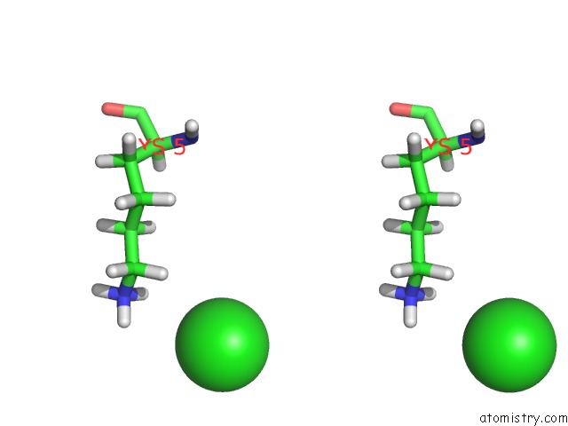
Stereo pair view
|
A full contact list of Chlorine with other atoms in the Cl binding
site number 10 of 1H-uc(Nmr) Solution Structure of A Trypsin/Chymotrypsin Bowman- Birk Inhibitor From Lens Culinaris. within 5.0Å range:
probe
|
atom
|
residue
|
distance (Å)
|
B
|
Occ
|
A:Cl77
b:0.0
occ:1.00
|
HZ2
|
A:LYS5
|
3.5
|
0.0
|
1.0
|
HZ1
|
A:LYS5
|
3.8
|
0.0
|
1.0
|
HE2
|
A:LYS5
|
3.9
|
0.0
|
1.0
|
NZ
|
A:LYS5
|
4.1
|
0.0
|
1.0
|
CE
|
A:LYS5
|
4.6
|
0.0
|
1.0
|
|
Reference:
E.M.Ragg,
V.Galbusera,
A.Scarafoni,
A.Negri,
G.Tedeschi,
A.Consonni,
F.Sessa,
M.Duranti.
Inhibitory Properties and Solution Structure of A Potent Bowman-Birk Protease Inhibitor From Lentil (Lens Culinaris, L) Seeds. Febs J. V. 273 4024 2006.
ISSN: ISSN 1742-464X
PubMed: 16889634
DOI: 10.1111/J.1742-4658.2006.05406.X
Page generated: Sat Jul 20 05:14:54 2024
|