Chlorine »
PDB 2ajf-2axw »
2aof »
Chlorine in PDB 2aof: Crystal Structure Analysis of Hiv-1 Protease Mutant V82A with A Substrate Analog P1-P6
Enzymatic activity of Crystal Structure Analysis of Hiv-1 Protease Mutant V82A with A Substrate Analog P1-P6
All present enzymatic activity of Crystal Structure Analysis of Hiv-1 Protease Mutant V82A with A Substrate Analog P1-P6:
3.4.23.16;
3.4.23.16;
Protein crystallography data
The structure of Crystal Structure Analysis of Hiv-1 Protease Mutant V82A with A Substrate Analog P1-P6, PDB code: 2aof
was solved by
Y.Tie,
P.I.Boross,
Y.F.Wang,
L.Gaddis,
F.Liu,
X.Chen,
J.Tozser,
R.W.Harrison,
I.T.Weber,
with X-Ray Crystallography technique. A brief refinement statistics is given in the table below:
| Resolution Low / High (Å) | 10.00 / 1.32 |
| Space group | P 21 21 2 |
| Cell size a, b, c (Å), α, β, γ (°) | 58.457, 85.851, 46.394, 90.00, 90.00, 90.00 |
| R / Rfree (%) | 13.1 / 18.4 |
Other elements in 2aof:
The structure of Crystal Structure Analysis of Hiv-1 Protease Mutant V82A with A Substrate Analog P1-P6 also contains other interesting chemical elements:
| Sodium | (Na) | 1 atom |
Chlorine Binding Sites:
The binding sites of Chlorine atom in the Crystal Structure Analysis of Hiv-1 Protease Mutant V82A with A Substrate Analog P1-P6
(pdb code 2aof). This binding sites where shown within
5.0 Angstroms radius around Chlorine atom.
In total 2 binding sites of Chlorine where determined in the Crystal Structure Analysis of Hiv-1 Protease Mutant V82A with A Substrate Analog P1-P6, PDB code: 2aof:
Jump to Chlorine binding site number: 1; 2;
In total 2 binding sites of Chlorine where determined in the Crystal Structure Analysis of Hiv-1 Protease Mutant V82A with A Substrate Analog P1-P6, PDB code: 2aof:
Jump to Chlorine binding site number: 1; 2;
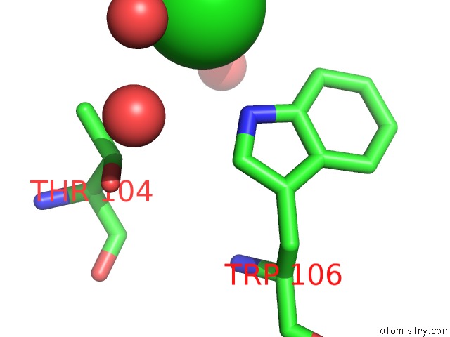
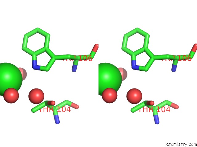
Chlorine binding site 1 out of 2 in 2aof
Go back to
Chlorine binding site 1 out
of 2 in the Crystal Structure Analysis of Hiv-1 Protease Mutant V82A with A Substrate Analog P1-P6

Mono view

Stereo pair view
Mono view
Stereo pair view
A full contact list of Chlorine with other atoms in the Cl binding
site number 1 of Crystal Structure Analysis of Hiv-1 Protease Mutant V82A with A Substrate Analog P1-P6 within 5.0Å range:
|
Chlorine binding site 2 out of 2 in 2aof
Go back to
Chlorine binding site 2 out
of 2 in the Crystal Structure Analysis of Hiv-1 Protease Mutant V82A with A Substrate Analog P1-P6
Mono view
Stereo pair view
Mono view
Stereo pair view
A full contact list of Chlorine with other atoms in the Cl binding
site number 2 of Crystal Structure Analysis of Hiv-1 Protease Mutant V82A with A Substrate Analog P1-P6 within 5.0Å range:
|
Reference:
Y.Tie,
P.I.Boross,
Y.F.Wang,
L.Gaddis,
F.Liu,
X.Chen,
J.Tozser,
R.W.Harrison,
I.T.Weber.
Molecular Basis For Substrate Recognition and Drug Resistance From 1.1 to 1.6 Angstroms Resolution Crystal Structures of Hiv-1 Protease Mutants with Substrate Analogs. Febs J. V. 272 5265 2005.
ISSN: ISSN 1742-464X
PubMed: 16218957
DOI: 10.1111/J.1742-4658.2005.04923.X
Page generated: Sat Jul 20 05:20:02 2024
ISSN: ISSN 1742-464X
PubMed: 16218957
DOI: 10.1111/J.1742-4658.2005.04923.X
Last articles
Zn in 9J0NZn in 9J0O
Zn in 9J0P
Zn in 9FJX
Zn in 9EKB
Zn in 9C0F
Zn in 9CAH
Zn in 9CH0
Zn in 9CH3
Zn in 9CH1