Chlorine »
PDB 2ay0-2bch »
2ay0 »
Chlorine in PDB 2ay0: Structure of the LYS9MET Mutant of the E. Coli Proline Utilization A (Puta) Dna-Binding Domain.
Protein crystallography data
The structure of Structure of the LYS9MET Mutant of the E. Coli Proline Utilization A (Puta) Dna-Binding Domain., PDB code: 2ay0
was solved by
J.D.Larson,
J.P.Schuermann,
Y.Zhou,
J.L.Jenkins,
D.F.Becker,
J.J.Tanner,
with X-Ray Crystallography technique. A brief refinement statistics is given in the table below:
| Resolution Low / High (Å) | 60.75 / 2.10 |
| Space group | C 1 2 1 |
| Cell size a, b, c (Å), α, β, γ (°) | 72.070, 91.494, 69.606, 90.00, 119.21, 90.00 |
| R / Rfree (%) | 20.4 / 24.2 |
Chlorine Binding Sites:
The binding sites of Chlorine atom in the Structure of the LYS9MET Mutant of the E. Coli Proline Utilization A (Puta) Dna-Binding Domain.
(pdb code 2ay0). This binding sites where shown within
5.0 Angstroms radius around Chlorine atom.
In total 6 binding sites of Chlorine where determined in the Structure of the LYS9MET Mutant of the E. Coli Proline Utilization A (Puta) Dna-Binding Domain., PDB code: 2ay0:
Jump to Chlorine binding site number: 1; 2; 3; 4; 5; 6;
In total 6 binding sites of Chlorine where determined in the Structure of the LYS9MET Mutant of the E. Coli Proline Utilization A (Puta) Dna-Binding Domain., PDB code: 2ay0:
Jump to Chlorine binding site number: 1; 2; 3; 4; 5; 6;
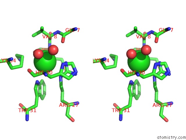
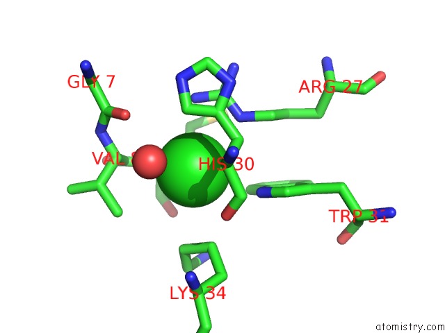
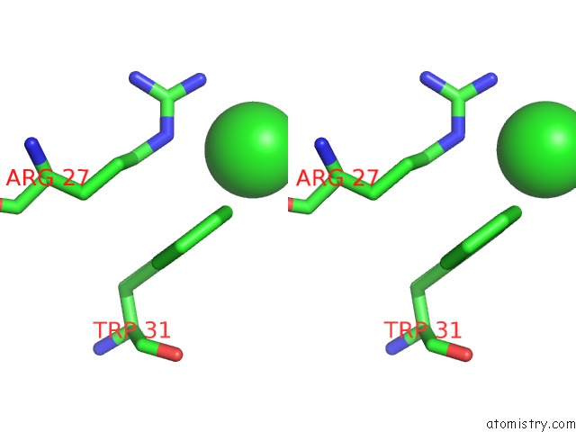
Chlorine binding site 1 out of 6 in 2ay0
Go back to
Chlorine binding site 1 out
of 6 in the Structure of the LYS9MET Mutant of the E. Coli Proline Utilization A (Puta) Dna-Binding Domain.

Mono view

Stereo pair view
Mono view
Stereo pair view
A full contact list of Chlorine with other atoms in the Cl binding
site number 1 of Structure of the LYS9MET Mutant of the E. Coli Proline Utilization A (Puta) Dna-Binding Domain. within 5.0Å range:
|
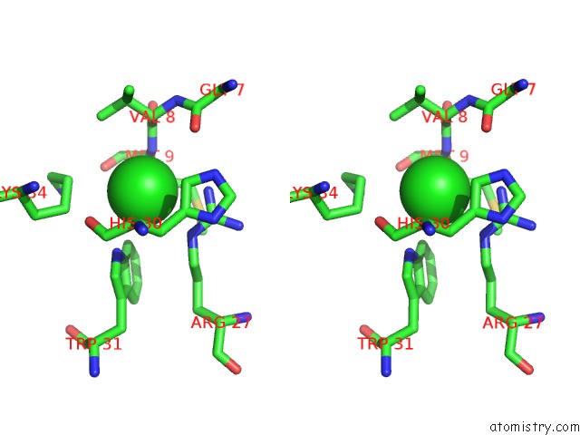
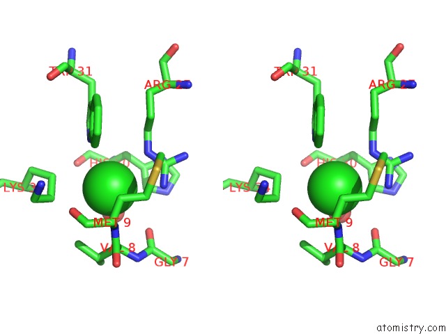
Chlorine binding site 2 out of 6 in 2ay0
Go back to
Chlorine binding site 2 out
of 6 in the Structure of the LYS9MET Mutant of the E. Coli Proline Utilization A (Puta) Dna-Binding Domain.

Mono view
Stereo pair view
Mono view
Stereo pair view
A full contact list of Chlorine with other atoms in the Cl binding
site number 2 of Structure of the LYS9MET Mutant of the E. Coli Proline Utilization A (Puta) Dna-Binding Domain. within 5.0Å range:
|
Chlorine binding site 3 out of 6 in 2ay0
Go back to
Chlorine binding site 3 out
of 6 in the Structure of the LYS9MET Mutant of the E. Coli Proline Utilization A (Puta) Dna-Binding Domain.
Mono view

Stereo pair view
Mono view
Stereo pair view
A full contact list of Chlorine with other atoms in the Cl binding
site number 3 of Structure of the LYS9MET Mutant of the E. Coli Proline Utilization A (Puta) Dna-Binding Domain. within 5.0Å range:
|
Chlorine binding site 4 out of 6 in 2ay0
Go back to
Chlorine binding site 4 out
of 6 in the Structure of the LYS9MET Mutant of the E. Coli Proline Utilization A (Puta) Dna-Binding Domain.

Mono view
Stereo pair view
Mono view
Stereo pair view
A full contact list of Chlorine with other atoms in the Cl binding
site number 4 of Structure of the LYS9MET Mutant of the E. Coli Proline Utilization A (Puta) Dna-Binding Domain. within 5.0Å range:
|
Chlorine binding site 5 out of 6 in 2ay0
Go back to
Chlorine binding site 5 out
of 6 in the Structure of the LYS9MET Mutant of the E. Coli Proline Utilization A (Puta) Dna-Binding Domain.

Mono view
Stereo pair view
Mono view
Stereo pair view
A full contact list of Chlorine with other atoms in the Cl binding
site number 5 of Structure of the LYS9MET Mutant of the E. Coli Proline Utilization A (Puta) Dna-Binding Domain. within 5.0Å range:
|
Chlorine binding site 6 out of 6 in 2ay0
Go back to
Chlorine binding site 6 out
of 6 in the Structure of the LYS9MET Mutant of the E. Coli Proline Utilization A (Puta) Dna-Binding Domain.

Mono view
Stereo pair view
Mono view
Stereo pair view
A full contact list of Chlorine with other atoms in the Cl binding
site number 6 of Structure of the LYS9MET Mutant of the E. Coli Proline Utilization A (Puta) Dna-Binding Domain. within 5.0Å range:
|
Reference:
J.D.Larson,
J.L.Jenkins,
J.P.Schuermann,
Y.Zhou,
D.F.Becker,
J.J.Tanner.
Crystal Structures of the Dna-Binding Domain of Escherichia Coli Proline Utilization A Flavoprotein and Analysis of the Role of LYS9 in Dna Recognition. Protein Sci. V. 15 2630 2006.
ISSN: ISSN 0961-8368
PubMed: 17001030
DOI: 10.1110/PS.062425706
Page generated: Thu Jul 10 21:17:52 2025
ISSN: ISSN 0961-8368
PubMed: 17001030
DOI: 10.1110/PS.062425706
Last articles
Fe in 2IAGFe in 2IAH
Fe in 2IA8
Fe in 2I9U
Fe in 2HTN
Fe in 2I96
Fe in 2I8F
Fe in 2I5N
Fe in 2I7F
Fe in 2HZ3