Chlorine »
PDB 2bl9-2bws »
2boh »
Chlorine in PDB 2boh: Crystal Structure of Factor Xa in Complex with Compound "1"
Enzymatic activity of Crystal Structure of Factor Xa in Complex with Compound "1"
All present enzymatic activity of Crystal Structure of Factor Xa in Complex with Compound "1":
3.4.21.6;
3.4.21.6;
Protein crystallography data
The structure of Crystal Structure of Factor Xa in Complex with Compound "1", PDB code: 2boh
was solved by
M.Nazare,
D.W.Will,
H.Matter,
H.Schreuder,
K.Ritter,
M.Urmann,
M.Essrich,
A.Bauer,
M.Wagner,
J.Czech,
V.Laux,
V.Wehner,
with X-Ray Crystallography technique. A brief refinement statistics is given in the table below:
| Resolution Low / High (Å) | 50.00 / 2.20 |
| Space group | P 21 21 21 |
| Cell size a, b, c (Å), α, β, γ (°) | 56.100, 72.200, 79.200, 90.00, 90.00, 90.00 |
| R / Rfree (%) | n/a / n/a |
Other elements in 2boh:
The structure of Crystal Structure of Factor Xa in Complex with Compound "1" also contains other interesting chemical elements:
| Calcium | (Ca) | 1 atom |
Chlorine Binding Sites:
The binding sites of Chlorine atom in the Crystal Structure of Factor Xa in Complex with Compound "1"
(pdb code 2boh). This binding sites where shown within
5.0 Angstroms radius around Chlorine atom.
In total only one binding site of Chlorine was determined in the Crystal Structure of Factor Xa in Complex with Compound "1", PDB code: 2boh:
In total only one binding site of Chlorine was determined in the Crystal Structure of Factor Xa in Complex with Compound "1", PDB code: 2boh:
Chlorine binding site 1 out of 1 in 2boh
Go back to
Chlorine binding site 1 out
of 1 in the Crystal Structure of Factor Xa in Complex with Compound "1"
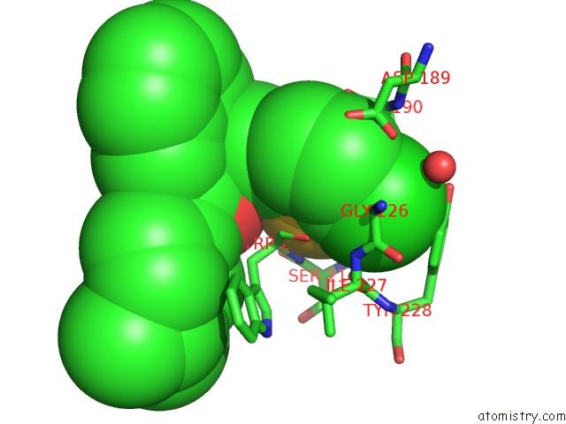
Mono view
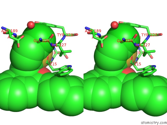
Stereo pair view
Mono view
Stereo pair view
A full contact list of Chlorine with other atoms in the Cl binding
site number 1 of Crystal Structure of Factor Xa in Complex with Compound "1" within 5.0Å range:
|
Reference:
M.Nazare,
D.W.Will,
H.Matter,
H.Schreuder,
K.Ritter,
M.Urmann,
M.Essrich,
A.Bauer,
M.Wagner,
J.Czech,
M.Lorenz,
V.Laux,
V.Wehner.
Probing the Subpockets of Factor Xa Reveals Two Binding Modes For Inhibitors Based on A 2-Carboxyindole Scaffold: A Study Combining Structure-Activity Relationship and X-Ray Crystallography. J.Med.Chem. V. 48 4511 2005.
ISSN: ISSN 0022-2623
PubMed: 15999990
DOI: 10.1021/JM0490540
Page generated: Sat Jul 20 05:50:27 2024
ISSN: ISSN 0022-2623
PubMed: 15999990
DOI: 10.1021/JM0490540
Last articles
Zn in 9JYWZn in 9IR4
Zn in 9IR3
Zn in 9GMX
Zn in 9GMW
Zn in 9JEJ
Zn in 9ERF
Zn in 9ERE
Zn in 9EGV
Zn in 9EGW