Chlorine »
PDB 2bla-2bwv »
2bu7 »
Chlorine in PDB 2bu7: Crystal Structures of Human Pyruvate Dehydrogenase Kinase 2 Containing Physiological and Synthetic Ligands
Enzymatic activity of Crystal Structures of Human Pyruvate Dehydrogenase Kinase 2 Containing Physiological and Synthetic Ligands
All present enzymatic activity of Crystal Structures of Human Pyruvate Dehydrogenase Kinase 2 Containing Physiological and Synthetic Ligands:
2.7.1.99;
2.7.1.99;
Protein crystallography data
The structure of Crystal Structures of Human Pyruvate Dehydrogenase Kinase 2 Containing Physiological and Synthetic Ligands, PDB code: 2bu7
was solved by
T.R.Knoechel,
A.D.Tucker,
C.M.Robinson,
C.Phillips,
W.Taylor,
P.J.Bungay,
S.A.Kasten,
T.E.Roche,
D.G.Brown,
with X-Ray Crystallography technique. A brief refinement statistics is given in the table below:
| Resolution Low / High (Å) | 30.00 / 2.40 |
| Space group | P 64 |
| Cell size a, b, c (Å), α, β, γ (°) | 108.876, 108.876, 84.025, 90.00, 90.00, 120.00 |
| R / Rfree (%) | n/a / n/a |
Chlorine Binding Sites:
The binding sites of Chlorine atom in the Crystal Structures of Human Pyruvate Dehydrogenase Kinase 2 Containing Physiological and Synthetic Ligands
(pdb code 2bu7). This binding sites where shown within
5.0 Angstroms radius around Chlorine atom.
In total only one binding site of Chlorine was determined in the Crystal Structures of Human Pyruvate Dehydrogenase Kinase 2 Containing Physiological and Synthetic Ligands, PDB code: 2bu7:
In total only one binding site of Chlorine was determined in the Crystal Structures of Human Pyruvate Dehydrogenase Kinase 2 Containing Physiological and Synthetic Ligands, PDB code: 2bu7:
Chlorine binding site 1 out of 1 in 2bu7
Go back to
Chlorine binding site 1 out
of 1 in the Crystal Structures of Human Pyruvate Dehydrogenase Kinase 2 Containing Physiological and Synthetic Ligands
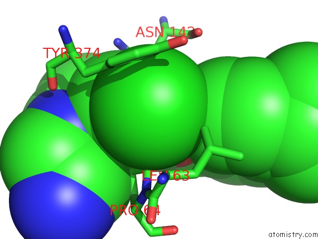
Mono view
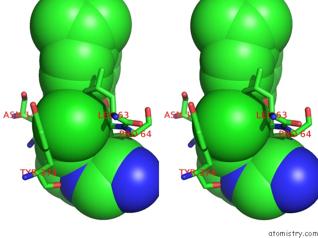
Stereo pair view
Mono view
Stereo pair view
A full contact list of Chlorine with other atoms in the Cl binding
site number 1 of Crystal Structures of Human Pyruvate Dehydrogenase Kinase 2 Containing Physiological and Synthetic Ligands within 5.0Å range:
|
Reference:
T.R.Knoechel,
A.D.Tucker,
C.M.Robinson,
C.Phillips,
W.Taylor,
P.J.Bungay,
S.A.Kasten,
T.E.Roche,
D.G.Brown.
Regulatory Roles of the N-Terminal Domain Based on Crystal Structures of Human Pyruvate Dehydrogenase Kinase 2 Containing Physiological and Synthetic Ligands. Biochemistry V. 45 402 2006.
ISSN: ISSN 0006-2960
PubMed: 16401071
DOI: 10.1021/BI051402S
Page generated: Sat Jul 20 05:54:50 2024
ISSN: ISSN 0006-2960
PubMed: 16401071
DOI: 10.1021/BI051402S
Last articles
Zn in 9MJ5Zn in 9HNW
Zn in 9G0L
Zn in 9FNE
Zn in 9DZN
Zn in 9E0I
Zn in 9D32
Zn in 9DAK
Zn in 8ZXC
Zn in 8ZUF