Chlorine »
PDB 2cie-2dch »
2cv5 »
Chlorine in PDB 2cv5: Crystal Structure of Human Nucleosome Core Particle
Protein crystallography data
The structure of Crystal Structure of Human Nucleosome Core Particle, PDB code: 2cv5
was solved by
Y.Tsunaka,
N.Kajimura,
S.Tate,
K.Morikawa,
with X-Ray Crystallography technique. A brief refinement statistics is given in the table below:
| Resolution Low / High (Å) | 50.00 / 2.50 |
| Space group | P 21 21 21 |
| Cell size a, b, c (Å), α, β, γ (°) | 99.564, 108.370, 169.425, 90.00, 90.00, 90.00 |
| R / Rfree (%) | 22.4 / 27.7 |
Other elements in 2cv5:
The structure of Crystal Structure of Human Nucleosome Core Particle also contains other interesting chemical elements:
| Manganese | (Mn) | 9 atoms |
Chlorine Binding Sites:
The binding sites of Chlorine atom in the Crystal Structure of Human Nucleosome Core Particle
(pdb code 2cv5). This binding sites where shown within
5.0 Angstroms radius around Chlorine atom.
In total 4 binding sites of Chlorine where determined in the Crystal Structure of Human Nucleosome Core Particle, PDB code: 2cv5:
Jump to Chlorine binding site number: 1; 2; 3; 4;
In total 4 binding sites of Chlorine where determined in the Crystal Structure of Human Nucleosome Core Particle, PDB code: 2cv5:
Jump to Chlorine binding site number: 1; 2; 3; 4;
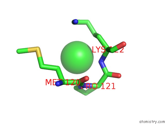
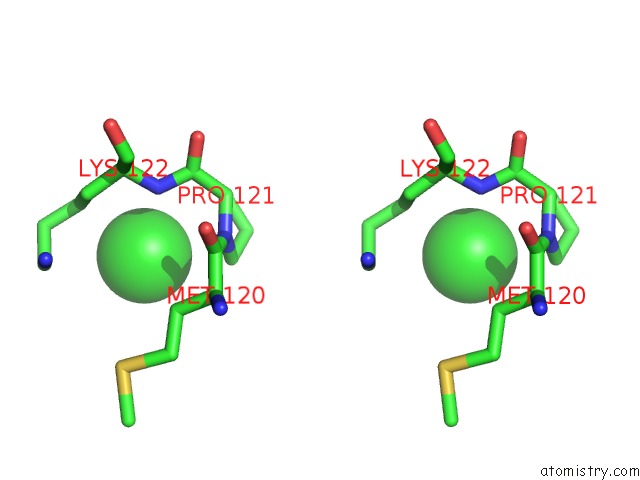
Chlorine binding site 1 out of 4 in 2cv5
Go back to
Chlorine binding site 1 out
of 4 in the Crystal Structure of Human Nucleosome Core Particle
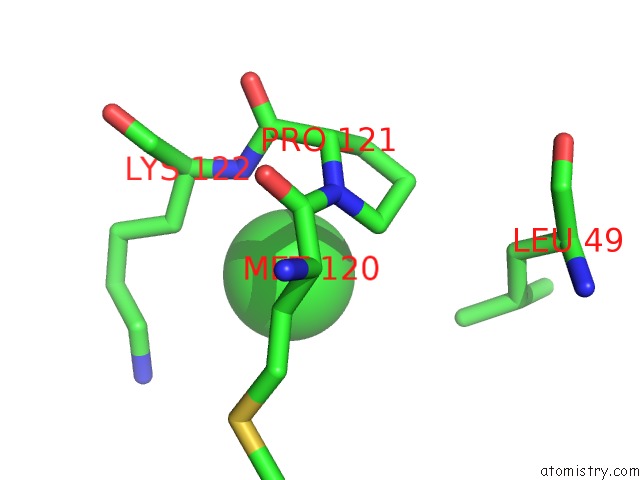
Mono view
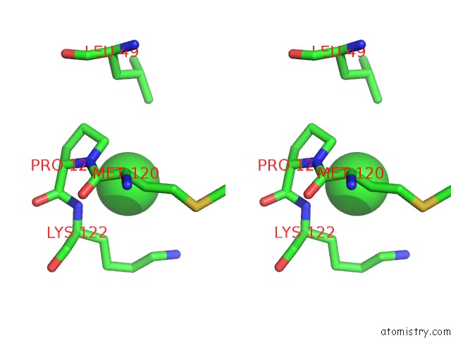
Stereo pair view
Mono view
Stereo pair view
A full contact list of Chlorine with other atoms in the Cl binding
site number 1 of Crystal Structure of Human Nucleosome Core Particle within 5.0Å range:
|
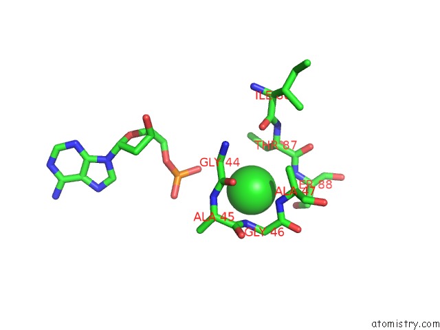
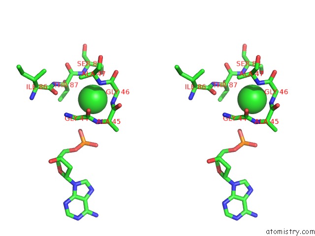
Chlorine binding site 2 out of 4 in 2cv5
Go back to
Chlorine binding site 2 out
of 4 in the Crystal Structure of Human Nucleosome Core Particle
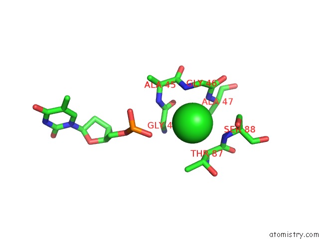
Mono view
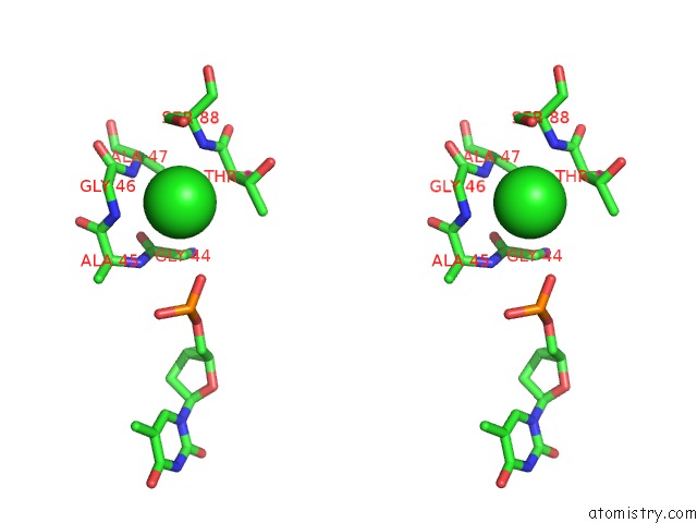
Stereo pair view
Mono view
Stereo pair view
A full contact list of Chlorine with other atoms in the Cl binding
site number 2 of Crystal Structure of Human Nucleosome Core Particle within 5.0Å range:
|
Chlorine binding site 3 out of 4 in 2cv5
Go back to
Chlorine binding site 3 out
of 4 in the Crystal Structure of Human Nucleosome Core Particle
Mono view
Stereo pair view
Mono view
Stereo pair view
A full contact list of Chlorine with other atoms in the Cl binding
site number 3 of Crystal Structure of Human Nucleosome Core Particle within 5.0Å range:
|
Chlorine binding site 4 out of 4 in 2cv5
Go back to
Chlorine binding site 4 out
of 4 in the Crystal Structure of Human Nucleosome Core Particle
Mono view
Stereo pair view
Mono view
Stereo pair view
A full contact list of Chlorine with other atoms in the Cl binding
site number 4 of Crystal Structure of Human Nucleosome Core Particle within 5.0Å range:
|
Reference:
Y.Tsunaka,
N.Kajimura,
S.Tate,
K.Morikawa.
Alteration of the Nucleosomal Dna Path in the Crystal Structure of A Human Nucleosome Core Particle Nucleic Acids Res. V. 33 3424 2005.
ISSN: ISSN 0305-1048
PubMed: 15951514
DOI: 10.1093/NAR/GKI663
Page generated: Sat Jul 20 06:24:16 2024
ISSN: ISSN 0305-1048
PubMed: 15951514
DOI: 10.1093/NAR/GKI663
Last articles
Zn in 9J0NZn in 9J0O
Zn in 9J0P
Zn in 9FJX
Zn in 9EKB
Zn in 9C0F
Zn in 9CAH
Zn in 9CH0
Zn in 9CH3
Zn in 9CH1