Chlorine in PDB 2dfd: Crystal Structure of Human Malate Dehydrogenase Type 2
Enzymatic activity of Crystal Structure of Human Malate Dehydrogenase Type 2
All present enzymatic activity of Crystal Structure of Human Malate Dehydrogenase Type 2:
1.1.1.37;
Protein crystallography data
The structure of Crystal Structure of Human Malate Dehydrogenase Type 2, PDB code: 2dfd
was solved by
E.Ugochukwu,
N.Shafqat,
A.Rojkova,
M.Sundstrom,
C.Arrowsmith,
J.Weigelt,
A.Edwards,
F.Von Delft,
U.Oppermann,
Structural Genomics Consortium(Sgc),
with X-Ray Crystallography technique. A brief refinement statistics is given in the table below:
Resolution Low / High (Å)
|
6.01 /
1.90
|
Space group
|
P 21 21 21
|
Cell size a, b, c (Å), α, β, γ (°)
|
59.943,
152.526,
154.827,
90.00,
90.00,
90.00
|
R / Rfree (%)
|
15.6 /
19.4
|
Chlorine Binding Sites:
The binding sites of Chlorine atom in the Crystal Structure of Human Malate Dehydrogenase Type 2
(pdb code 2dfd). This binding sites where shown within
5.0 Angstroms radius around Chlorine atom.
In total 7 binding sites of Chlorine where determined in the
Crystal Structure of Human Malate Dehydrogenase Type 2, PDB code: 2dfd:
Jump to Chlorine binding site number:
1;
2;
3;
4;
5;
6;
7;
Chlorine binding site 1 out
of 7 in 2dfd
Go back to
Chlorine Binding Sites List in 2dfd
Chlorine binding site 1 out
of 7 in the Crystal Structure of Human Malate Dehydrogenase Type 2
 Mono view
 Stereo pair view
|
A full contact list of Chlorine with other atoms in the Cl binding
site number 1 of Crystal Structure of Human Malate Dehydrogenase Type 2 within 5.0Å range:
probe
|
atom
|
residue
|
distance (Å)
|
B
|
Occ
|
A:Cl3203
b:36.3
occ:1.00
|
NH1
|
A:ARG92
|
3.1
|
37.7
|
1.0
|
O
|
A:HOH3343
|
3.1
|
33.5
|
1.0
|
O
|
A:HOH3398
|
3.2
|
41.5
|
1.0
|
N
|
A:ARG92
|
3.2
|
32.4
|
1.0
|
CG
|
A:ARG92
|
3.5
|
33.0
|
1.0
|
CB
|
A:ARG92
|
3.7
|
33.2
|
1.0
|
CD
|
A:ARG92
|
3.9
|
35.1
|
1.0
|
CA
|
A:ARG92
|
4.0
|
32.9
|
1.0
|
CB
|
A:ALA183
|
4.1
|
34.9
|
1.0
|
CA
|
A:THR91
|
4.1
|
32.2
|
1.0
|
C
|
A:THR91
|
4.1
|
31.9
|
1.0
|
CZ
|
A:ARG92
|
4.2
|
37.3
|
1.0
|
CB
|
A:THR91
|
4.2
|
33.3
|
1.0
|
NH1
|
A:ARG86
|
4.4
|
31.7
|
1.0
|
NE
|
A:ARG92
|
4.5
|
35.7
|
1.0
|
O
|
A:HOH3351
|
4.5
|
34.3
|
1.0
|
CA
|
A:ALA183
|
4.6
|
35.8
|
1.0
|
N
|
A:GLY184
|
4.6
|
35.1
|
1.0
|
CZ
|
A:ARG86
|
4.7
|
32.4
|
1.0
|
O
|
A:HOH3393
|
4.8
|
43.4
|
1.0
|
N
|
A:ASP93
|
4.9
|
33.5
|
1.0
|
NH2
|
A:ARG86
|
5.0
|
33.4
|
1.0
|
C
|
A:ALA183
|
5.0
|
35.8
|
1.0
|
|
Chlorine binding site 2 out
of 7 in 2dfd
Go back to
Chlorine Binding Sites List in 2dfd
Chlorine binding site 2 out
of 7 in the Crystal Structure of Human Malate Dehydrogenase Type 2
 Mono view
 Stereo pair view
|
A full contact list of Chlorine with other atoms in the Cl binding
site number 2 of Crystal Structure of Human Malate Dehydrogenase Type 2 within 5.0Å range:
probe
|
atom
|
residue
|
distance (Å)
|
B
|
Occ
|
A:Cl3205
b:86.3
occ:1.00
|
ND2
|
A:ASN146
|
3.1
|
34.2
|
1.0
|
O
|
A:HOH3399
|
3.6
|
52.5
|
1.0
|
O
|
A:HOH3363
|
3.7
|
30.1
|
1.0
|
O
|
A:PRO145
|
3.9
|
30.6
|
1.0
|
O
|
A:LYS261
|
3.9
|
29.7
|
1.0
|
CB
|
A:PRO145
|
3.9
|
30.1
|
1.0
|
C
|
A:PRO145
|
4.0
|
30.1
|
1.0
|
CG
|
A:PRO145
|
4.2
|
31.9
|
1.0
|
CD1
|
A:ILE281
|
4.3
|
30.7
|
1.0
|
CG
|
A:ASN146
|
4.3
|
33.7
|
1.0
|
N
|
A:ASN146
|
4.3
|
29.7
|
1.0
|
CB
|
A:LYS261
|
4.6
|
30.9
|
1.0
|
CA
|
A:ASN146
|
4.6
|
30.1
|
1.0
|
CA
|
A:PRO145
|
4.6
|
30.4
|
1.0
|
O
|
A:HOH3465
|
4.7
|
52.0
|
1.0
|
C
|
A:LYS261
|
4.7
|
31.1
|
1.0
|
CG
|
A:LYS261
|
4.8
|
31.5
|
1.0
|
|
Chlorine binding site 3 out
of 7 in 2dfd
Go back to
Chlorine Binding Sites List in 2dfd
Chlorine binding site 3 out
of 7 in the Crystal Structure of Human Malate Dehydrogenase Type 2
 Mono view
 Stereo pair view
|
A full contact list of Chlorine with other atoms in the Cl binding
site number 3 of Crystal Structure of Human Malate Dehydrogenase Type 2 within 5.0Å range:
probe
|
atom
|
residue
|
distance (Å)
|
B
|
Occ
|
B:Cl3206
b:76.5
occ:1.00
|
O
|
B:HOH3533
|
2.6
|
56.8
|
1.0
|
O
|
B:VAL198
|
3.1
|
36.2
|
1.0
|
N
|
B:PHE200
|
3.5
|
35.6
|
1.0
|
O
|
B:PHE200
|
4.1
|
34.4
|
1.0
|
O
|
B:HOH3520
|
4.1
|
54.6
|
1.0
|
CA
|
B:ASP199
|
4.1
|
36.5
|
1.0
|
CG2
|
B:ILE191
|
4.2
|
32.1
|
1.0
|
C
|
B:VAL198
|
4.2
|
35.6
|
1.0
|
CB
|
B:PHE200
|
4.3
|
35.3
|
1.0
|
C
|
B:ASP199
|
4.3
|
36.3
|
1.0
|
CD2
|
B:PHE200
|
4.3
|
35.6
|
1.0
|
CA
|
B:PHE200
|
4.4
|
35.4
|
1.0
|
CD2
|
B:LEU205
|
4.6
|
31.9
|
1.0
|
N
|
B:ASP199
|
4.6
|
35.8
|
1.0
|
C
|
B:PHE200
|
4.7
|
34.8
|
1.0
|
OD1
|
B:ASP199
|
4.8
|
41.6
|
1.0
|
CG
|
B:PHE200
|
4.8
|
34.8
|
1.0
|
CB
|
B:ILE191
|
4.8
|
31.9
|
1.0
|
O
|
B:ILE191
|
4.9
|
29.6
|
1.0
|
O
|
B:HOH3524
|
4.9
|
44.4
|
1.0
|
C
|
B:ILE191
|
5.0
|
30.8
|
1.0
|
|
Chlorine binding site 4 out
of 7 in 2dfd
Go back to
Chlorine Binding Sites List in 2dfd
Chlorine binding site 4 out
of 7 in the Crystal Structure of Human Malate Dehydrogenase Type 2
 Mono view
 Stereo pair view
|
A full contact list of Chlorine with other atoms in the Cl binding
site number 4 of Crystal Structure of Human Malate Dehydrogenase Type 2 within 5.0Å range:
probe
|
atom
|
residue
|
distance (Å)
|
B
|
Occ
|
C:Cl3201
b:28.7
occ:1.00
|
O
|
C:HOH3553
|
3.1
|
29.9
|
1.0
|
NH1
|
C:ARG92
|
3.2
|
32.3
|
1.0
|
N
|
C:ARG92
|
3.2
|
30.8
|
1.0
|
O
|
C:HOH3740
|
3.3
|
45.0
|
1.0
|
O
|
C:HOH3623
|
3.5
|
37.5
|
1.0
|
CG
|
C:ARG92
|
3.6
|
30.3
|
1.0
|
CB
|
C:ARG92
|
3.8
|
31.1
|
1.0
|
CB
|
C:ALA183
|
4.0
|
33.1
|
1.0
|
CD
|
C:ARG92
|
4.0
|
29.5
|
1.0
|
CA
|
C:THR91
|
4.1
|
31.9
|
1.0
|
CA
|
C:ARG92
|
4.1
|
31.1
|
1.0
|
CB
|
C:THR91
|
4.1
|
32.6
|
1.0
|
C
|
C:THR91
|
4.1
|
32.3
|
1.0
|
CZ
|
C:ARG92
|
4.3
|
33.8
|
1.0
|
NH1
|
C:ARG86
|
4.3
|
31.8
|
1.0
|
CA
|
C:ALA183
|
4.3
|
33.7
|
1.0
|
O
|
C:HOH3550
|
4.4
|
23.6
|
1.0
|
N
|
C:GLY184
|
4.4
|
33.9
|
1.0
|
NE
|
C:ARG92
|
4.6
|
32.3
|
1.0
|
O
|
C:HOH3584
|
4.6
|
32.2
|
1.0
|
CZ
|
C:ARG86
|
4.6
|
29.1
|
1.0
|
C
|
C:ALA183
|
4.7
|
34.0
|
1.0
|
CG2
|
C:THR91
|
4.8
|
32.2
|
1.0
|
NH2
|
C:ARG86
|
4.9
|
32.1
|
1.0
|
N
|
C:ASP93
|
4.9
|
31.1
|
1.0
|
|
Chlorine binding site 5 out
of 7 in 2dfd
Go back to
Chlorine Binding Sites List in 2dfd
Chlorine binding site 5 out
of 7 in the Crystal Structure of Human Malate Dehydrogenase Type 2
 Mono view
 Stereo pair view
|
A full contact list of Chlorine with other atoms in the Cl binding
site number 5 of Crystal Structure of Human Malate Dehydrogenase Type 2 within 5.0Å range:
probe
|
atom
|
residue
|
distance (Å)
|
B
|
Occ
|
C:Cl3204
b:69.4
occ:1.00
|
CG
|
C:GLN263
|
3.6
|
41.2
|
1.0
|
N
|
C:GLN263
|
3.7
|
34.6
|
1.0
|
C
|
C:SER262
|
3.7
|
33.7
|
1.0
|
NZ
|
C:LYS261
|
3.8
|
34.1
|
1.0
|
CB
|
C:GLN263
|
3.9
|
36.9
|
1.0
|
CA
|
C:SER262
|
4.0
|
32.8
|
1.0
|
N
|
C:SER262
|
4.1
|
31.2
|
1.0
|
CG
|
C:LYS261
|
4.1
|
32.7
|
1.0
|
O
|
C:SER262
|
4.2
|
33.8
|
1.0
|
CE
|
C:LYS261
|
4.3
|
38.1
|
1.0
|
O
|
C:HOH3731
|
4.3
|
43.5
|
1.0
|
CD
|
C:GLN263
|
4.4
|
42.4
|
1.0
|
C
|
C:LYS261
|
4.4
|
32.5
|
1.0
|
CA
|
C:GLN263
|
4.4
|
37.2
|
1.0
|
O
|
C:LYS261
|
4.5
|
31.7
|
1.0
|
OE1
|
C:GLN263
|
4.7
|
51.7
|
1.0
|
CD
|
C:LYS261
|
4.8
|
35.4
|
1.0
|
O
|
C:HOH3657
|
4.9
|
38.7
|
1.0
|
|
Chlorine binding site 6 out
of 7 in 2dfd
Go back to
Chlorine Binding Sites List in 2dfd
Chlorine binding site 6 out
of 7 in the Crystal Structure of Human Malate Dehydrogenase Type 2
 Mono view
 Stereo pair view
|
A full contact list of Chlorine with other atoms in the Cl binding
site number 6 of Crystal Structure of Human Malate Dehydrogenase Type 2 within 5.0Å range:
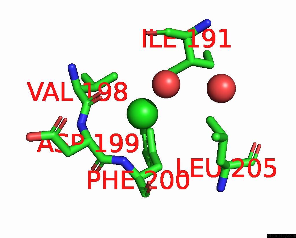
probe
|
atom
|
residue
|
distance (Å)
|
B
|
Occ
|
D:Cl3202
b:38.3
occ:1.00
|
O
|
D:HOH3318
|
2.9
|
35.7
|
1.0
|
NH1
|
D:ARG92
|
3.2
|
38.0
|
1.0
|
N
|
D:ARG92
|
3.3
|
33.5
|
1.0
|
O
|
D:HOH3403
|
3.4
|
48.8
|
1.0
|
CG
|
D:ARG92
|
3.7
|
32.9
|
1.0
|
CB
|
D:ARG92
|
3.8
|
32.0
|
1.0
|
CD
|
D:ARG92
|
3.9
|
34.8
|
1.0
|
CA
|
D:THR91
|
4.0
|
33.6
|
1.0
|
CB
|
D:ALA183
|
4.1
|
36.8
|
1.0
|
CB
|
D:THR91
|
4.1
|
33.5
|
1.0
|
C
|
D:THR91
|
4.1
|
32.6
|
1.0
|
CA
|
D:ARG92
|
4.1
|
33.0
|
1.0
|
CZ
|
D:ARG92
|
4.2
|
36.1
|
1.0
|
NH2
|
D:ARG86
|
4.3
|
32.4
|
1.0
|
CA
|
D:ALA183
|
4.5
|
35.9
|
1.0
|
NE
|
D:ARG92
|
4.5
|
37.1
|
1.0
|
O
|
D:HOH3402
|
4.6
|
33.2
|
1.0
|
N
|
D:GLY184
|
4.6
|
36.5
|
1.0
|
CZ
|
D:ARG86
|
4.7
|
33.3
|
1.0
|
C
|
D:ALA183
|
4.9
|
36.0
|
1.0
|
CG2
|
D:THR91
|
4.9
|
34.2
|
1.0
|
N
|
D:ASP93
|
5.0
|
34.3
|
1.0
|
|
Chlorine binding site 7 out
of 7 in 2dfd
Go back to
Chlorine Binding Sites List in 2dfd
Chlorine binding site 7 out
of 7 in the Crystal Structure of Human Malate Dehydrogenase Type 2
 Mono view
 Stereo pair view
|
A full contact list of Chlorine with other atoms in the Cl binding
site number 7 of Crystal Structure of Human Malate Dehydrogenase Type 2 within 5.0Å range:
probe
|
atom
|
residue
|
distance (Å)
|
B
|
Occ
|
D:Cl3207
b:77.7
occ:1.00
|
O
|
D:HOH3416
|
2.5
|
53.9
|
1.0
|
O
|
D:VAL198
|
3.5
|
35.9
|
1.0
|
N
|
D:PHE200
|
3.7
|
35.3
|
1.0
|
CA
|
D:ASP199
|
4.0
|
37.5
|
1.0
|
O
|
D:PHE200
|
4.1
|
34.6
|
1.0
|
CD2
|
D:LEU205
|
4.3
|
33.7
|
1.0
|
C
|
D:ASP199
|
4.4
|
36.4
|
1.0
|
OD1
|
D:ASP199
|
4.5
|
49.0
|
1.0
|
C
|
D:VAL198
|
4.5
|
36.5
|
1.0
|
CD1
|
D:LEU205
|
4.7
|
33.5
|
1.0
|
CG2
|
D:ILE191
|
4.7
|
32.5
|
1.0
|
CA
|
D:PHE200
|
4.7
|
34.5
|
1.0
|
N
|
D:ASP199
|
4.8
|
36.8
|
1.0
|
CB
|
D:PHE200
|
4.8
|
34.0
|
1.0
|
CD2
|
D:PHE200
|
4.8
|
32.2
|
1.0
|
C
|
D:PHE200
|
4.9
|
34.3
|
1.0
|
O
|
D:HOH3223
|
5.0
|
30.5
|
1.0
|
CB
|
D:ASP199
|
5.0
|
39.0
|
1.0
|
|
Reference:
E.Ugochukwu,
N.Shafqat,
A.Rojkova,
M.Sundstrom,
C.Arrowsmith,
J.Weigelt,
A.Edwards,
F.Von Delft,
U.Oppermann.
Crystal Structure of Human Malate Dehydrogenase Type 2 To Be Published.
Page generated: Sat Jul 20 06:31:32 2024
|