Chlorine »
PDB 2etd-2f81 »
2etl »
Chlorine in PDB 2etl: Crystal Structure of Ubiquitin Carboxy-Terminal Hydrolase L1 (Uch-L1)
Enzymatic activity of Crystal Structure of Ubiquitin Carboxy-Terminal Hydrolase L1 (Uch-L1)
All present enzymatic activity of Crystal Structure of Ubiquitin Carboxy-Terminal Hydrolase L1 (Uch-L1):
3.4.19.12;
3.4.19.12;
Protein crystallography data
The structure of Crystal Structure of Ubiquitin Carboxy-Terminal Hydrolase L1 (Uch-L1), PDB code: 2etl
was solved by
C.Das,
Q.Q.Hoang,
C.A.Kreinbring,
S.J.Luchansky,
R.K.Meray,
S.S.Ray,
P.T.Lansbury,
D.Ringe,
G.A.Petsko,
with X-Ray Crystallography technique. A brief refinement statistics is given in the table below:
| Resolution Low / High (Å) | 41.90 / 2.40 |
| Space group | P 4 21 2 |
| Cell size a, b, c (Å), α, β, γ (°) | 110.097, 110.097, 79.489, 90.00, 90.00, 90.00 |
| R / Rfree (%) | 22.3 / 27.4 |
Chlorine Binding Sites:
The binding sites of Chlorine atom in the Crystal Structure of Ubiquitin Carboxy-Terminal Hydrolase L1 (Uch-L1)
(pdb code 2etl). This binding sites where shown within
5.0 Angstroms radius around Chlorine atom.
In total 4 binding sites of Chlorine where determined in the Crystal Structure of Ubiquitin Carboxy-Terminal Hydrolase L1 (Uch-L1), PDB code: 2etl:
Jump to Chlorine binding site number: 1; 2; 3; 4;
In total 4 binding sites of Chlorine where determined in the Crystal Structure of Ubiquitin Carboxy-Terminal Hydrolase L1 (Uch-L1), PDB code: 2etl:
Jump to Chlorine binding site number: 1; 2; 3; 4;
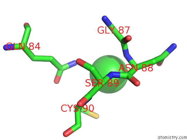
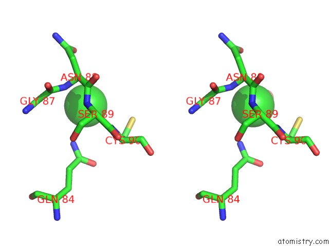
Chlorine binding site 1 out of 4 in 2etl
Go back to
Chlorine binding site 1 out
of 4 in the Crystal Structure of Ubiquitin Carboxy-Terminal Hydrolase L1 (Uch-L1)
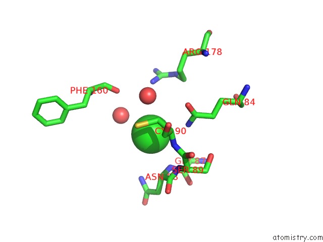
Mono view
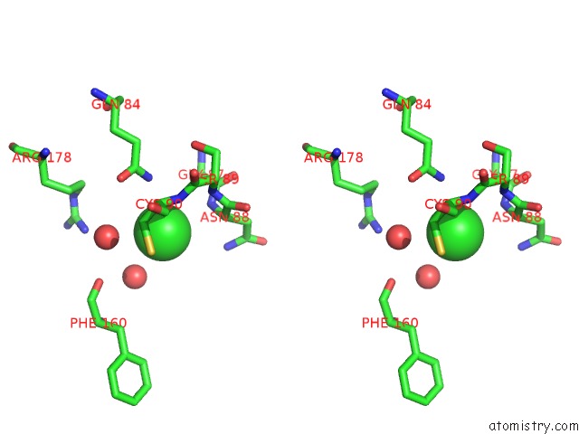
Stereo pair view
Mono view
Stereo pair view
A full contact list of Chlorine with other atoms in the Cl binding
site number 1 of Crystal Structure of Ubiquitin Carboxy-Terminal Hydrolase L1 (Uch-L1) within 5.0Å range:
|
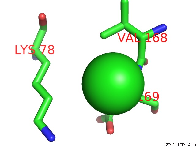
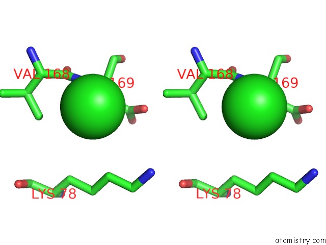
Chlorine binding site 2 out of 4 in 2etl
Go back to
Chlorine binding site 2 out
of 4 in the Crystal Structure of Ubiquitin Carboxy-Terminal Hydrolase L1 (Uch-L1)
Mono view
Stereo pair view
Mono view
Stereo pair view
A full contact list of Chlorine with other atoms in the Cl binding
site number 2 of Crystal Structure of Ubiquitin Carboxy-Terminal Hydrolase L1 (Uch-L1) within 5.0Å range:
|
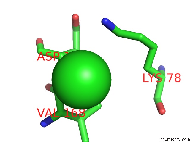
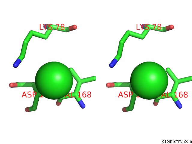
Chlorine binding site 3 out of 4 in 2etl
Go back to
Chlorine binding site 3 out
of 4 in the Crystal Structure of Ubiquitin Carboxy-Terminal Hydrolase L1 (Uch-L1)
Mono view
Stereo pair view
Mono view
Stereo pair view
A full contact list of Chlorine with other atoms in the Cl binding
site number 3 of Crystal Structure of Ubiquitin Carboxy-Terminal Hydrolase L1 (Uch-L1) within 5.0Å range:
|
Chlorine binding site 4 out of 4 in 2etl
Go back to
Chlorine binding site 4 out
of 4 in the Crystal Structure of Ubiquitin Carboxy-Terminal Hydrolase L1 (Uch-L1)
Mono view
Stereo pair view
Mono view
Stereo pair view
A full contact list of Chlorine with other atoms in the Cl binding
site number 4 of Crystal Structure of Ubiquitin Carboxy-Terminal Hydrolase L1 (Uch-L1) within 5.0Å range:
|
Reference:
C.Das,
Q.Q.Hoang,
C.A.Kreinbring,
S.J.Luchansky,
R.K.Meray,
S.S.Ray,
P.T.Lansbury,
D.Ringe,
G.A.Petsko.
Structural Basis For Conformational Plasticity of the Parkinson'S Disease-Associated Ubiquitin Hydrolase Uch-L1. Proc.Natl.Acad.Sci.Usa V. 103 4675 2006.
ISSN: ISSN 0027-8424
PubMed: 16537382
DOI: 10.1073/PNAS.0510403103
Page generated: Sat Jul 20 06:51:52 2024
ISSN: ISSN 0027-8424
PubMed: 16537382
DOI: 10.1073/PNAS.0510403103
Last articles
Zn in 9J0NZn in 9J0O
Zn in 9J0P
Zn in 9FJX
Zn in 9EKB
Zn in 9C0F
Zn in 9CAH
Zn in 9CH0
Zn in 9CH3
Zn in 9CH1