Chlorine »
PDB 2fs7-2g7z »
2fyq »
Chlorine in PDB 2fyq: Crystal Structure of the Norwalk Virus Protease
Protein crystallography data
The structure of Crystal Structure of the Norwalk Virus Protease, PDB code: 2fyq
was solved by
C.E.Zeitler,
M.K.Estes,
B.V.Venkataram Prasad,
with X-Ray Crystallography technique. A brief refinement statistics is given in the table below:
| Resolution Low / High (Å) | 105.41 / 1.50 |
| Space group | P 65 2 2 |
| Cell size a, b, c (Å), α, β, γ (°) | 121.833, 121.833, 51.369, 90.00, 90.00, 120.00 |
| R / Rfree (%) | 18.5 / 21.1 |
Chlorine Binding Sites:
The binding sites of Chlorine atom in the Crystal Structure of the Norwalk Virus Protease
(pdb code 2fyq). This binding sites where shown within
5.0 Angstroms radius around Chlorine atom.
In total 2 binding sites of Chlorine where determined in the Crystal Structure of the Norwalk Virus Protease, PDB code: 2fyq:
Jump to Chlorine binding site number: 1; 2;
In total 2 binding sites of Chlorine where determined in the Crystal Structure of the Norwalk Virus Protease, PDB code: 2fyq:
Jump to Chlorine binding site number: 1; 2;
Chlorine binding site 1 out of 2 in 2fyq
Go back to
Chlorine binding site 1 out
of 2 in the Crystal Structure of the Norwalk Virus Protease
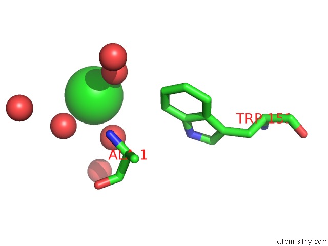
Mono view
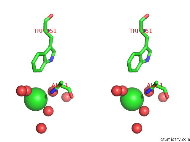
Stereo pair view
Mono view
Stereo pair view
A full contact list of Chlorine with other atoms in the Cl binding
site number 1 of Crystal Structure of the Norwalk Virus Protease within 5.0Å range:
|
Chlorine binding site 2 out of 2 in 2fyq
Go back to
Chlorine binding site 2 out
of 2 in the Crystal Structure of the Norwalk Virus Protease
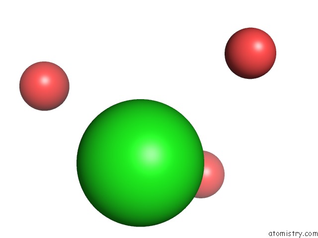
Mono view
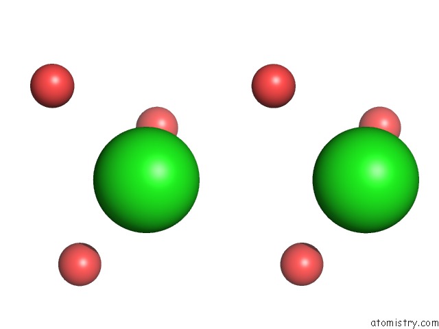
Stereo pair view
Mono view
Stereo pair view
A full contact list of Chlorine with other atoms in the Cl binding
site number 2 of Crystal Structure of the Norwalk Virus Protease within 5.0Å range:
|
Reference:
C.E.Zeitler,
M.K.Estes,
B.V.Venkataram Prasad.
X-Ray Crystallographic Structure of the Norwalk Virus Protease at 1.5-A Resolution. J.Virol. V. 80 5050 2006.
ISSN: ISSN 0022-538X
PubMed: 16641296
DOI: 10.1128/JVI.80.10.5050-5058.2006
Page generated: Sat Jul 20 07:14:56 2024
ISSN: ISSN 0022-538X
PubMed: 16641296
DOI: 10.1128/JVI.80.10.5050-5058.2006
Last articles
Zn in 9J0NZn in 9J0O
Zn in 9J0P
Zn in 9FJX
Zn in 9EKB
Zn in 9C0F
Zn in 9CAH
Zn in 9CH0
Zn in 9CH3
Zn in 9CH1