Chlorine »
PDB 2fs7-2g7z »
2g4w »
Chlorine in PDB 2g4w: Anomalous Substructure of Ribonuclease A (C2)
Enzymatic activity of Anomalous Substructure of Ribonuclease A (C2)
All present enzymatic activity of Anomalous Substructure of Ribonuclease A (C2):
3.1.27.5;
3.1.27.5;
Protein crystallography data
The structure of Anomalous Substructure of Ribonuclease A (C2), PDB code: 2g4w
was solved by
C.Mueller-Dieckmann,
M.S.Weiss,
with X-Ray Crystallography technique. A brief refinement statistics is given in the table below:
| Resolution Low / High (Å) | 30.00 / 1.84 |
| Space group | C 1 2 1 |
| Cell size a, b, c (Å), α, β, γ (°) | 100.120, 32.600, 72.470, 90.00, 90.56, 90.00 |
| R / Rfree (%) | 21.3 / 27.4 |
Chlorine Binding Sites:
The binding sites of Chlorine atom in the Anomalous Substructure of Ribonuclease A (C2)
(pdb code 2g4w). This binding sites where shown within
5.0 Angstroms radius around Chlorine atom.
In total only one binding site of Chlorine was determined in the Anomalous Substructure of Ribonuclease A (C2), PDB code: 2g4w:
In total only one binding site of Chlorine was determined in the Anomalous Substructure of Ribonuclease A (C2), PDB code: 2g4w:
Chlorine binding site 1 out of 1 in 2g4w
Go back to
Chlorine binding site 1 out
of 1 in the Anomalous Substructure of Ribonuclease A (C2)
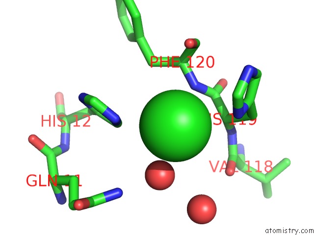
Mono view
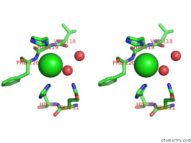
Stereo pair view
Mono view
Stereo pair view
A full contact list of Chlorine with other atoms in the Cl binding
site number 1 of Anomalous Substructure of Ribonuclease A (C2) within 5.0Å range:
|
Reference:
C.Mueller-Dieckmann,
S.Panjikar,
A.Schmidt,
S.Mueller,
J.Kuper,
A.Geerlof,
M.Wilmanns,
R.K.Singh,
P.A.Tucker,
M.S.Weiss.
On the Routine Use of Soft X-Rays in Macromolecular Crystallography. Part IV. Efficient Determination of Anomalous Substructures in Biomacromolecules Using Longer X-Ray Wavelengths. Acta Crystallogr.,Sect.D V. 63 366 2007.
ISSN: ISSN 0907-4449
PubMed: 17327674
DOI: 10.1107/S0907444906055624
Page generated: Sat Jul 20 07:20:15 2024
ISSN: ISSN 0907-4449
PubMed: 17327674
DOI: 10.1107/S0907444906055624
Last articles
Zn in 9J0NZn in 9J0O
Zn in 9J0P
Zn in 9FJX
Zn in 9EKB
Zn in 9C0F
Zn in 9CAH
Zn in 9CH0
Zn in 9CH3
Zn in 9CH1