Chlorine »
PDB 2fs7-2g7z »
2g7f »
Chlorine in PDB 2g7f: The 1.95 A Crystal Structure of Vibrio Cholerae Extracellular Endonuclease I
Enzymatic activity of The 1.95 A Crystal Structure of Vibrio Cholerae Extracellular Endonuclease I
All present enzymatic activity of The 1.95 A Crystal Structure of Vibrio Cholerae Extracellular Endonuclease I:
3.1.21.1;
3.1.21.1;
Protein crystallography data
The structure of The 1.95 A Crystal Structure of Vibrio Cholerae Extracellular Endonuclease I, PDB code: 2g7f
was solved by
B.Altermark,
A.O.Smalaas,
N.P.Willassen,
R.Helland,
with X-Ray Crystallography technique. A brief refinement statistics is given in the table below:
| Resolution Low / High (Å) | 35.65 / 1.95 |
| Space group | P 21 21 21 |
| Cell size a, b, c (Å), α, β, γ (°) | 40.400, 64.750, 75.780, 90.00, 90.00, 90.00 |
| R / Rfree (%) | 17.7 / 23.3 |
Other elements in 2g7f:
The structure of The 1.95 A Crystal Structure of Vibrio Cholerae Extracellular Endonuclease I also contains other interesting chemical elements:
| Magnesium | (Mg) | 1 atom |
Chlorine Binding Sites:
The binding sites of Chlorine atom in the The 1.95 A Crystal Structure of Vibrio Cholerae Extracellular Endonuclease I
(pdb code 2g7f). This binding sites where shown within
5.0 Angstroms radius around Chlorine atom.
In total only one binding site of Chlorine was determined in the The 1.95 A Crystal Structure of Vibrio Cholerae Extracellular Endonuclease I, PDB code: 2g7f:
In total only one binding site of Chlorine was determined in the The 1.95 A Crystal Structure of Vibrio Cholerae Extracellular Endonuclease I, PDB code: 2g7f:
Chlorine binding site 1 out of 1 in 2g7f
Go back to
Chlorine binding site 1 out
of 1 in the The 1.95 A Crystal Structure of Vibrio Cholerae Extracellular Endonuclease I
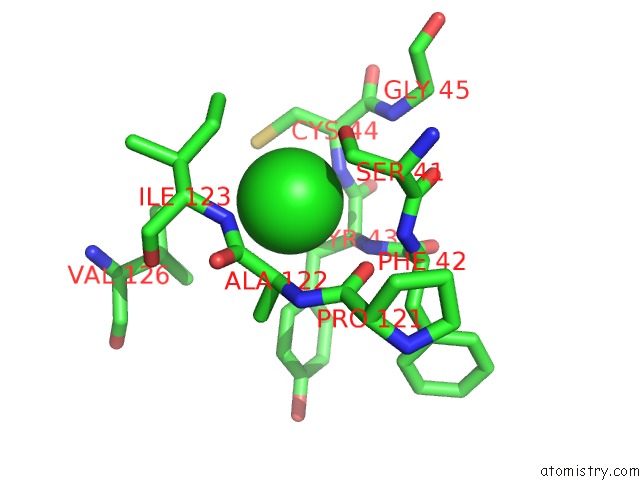
Mono view
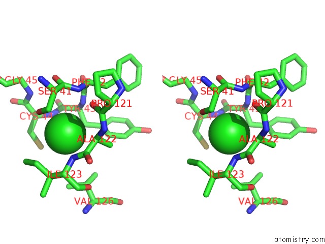
Stereo pair view
Mono view
Stereo pair view
A full contact list of Chlorine with other atoms in the Cl binding
site number 1 of The 1.95 A Crystal Structure of Vibrio Cholerae Extracellular Endonuclease I within 5.0Å range:
|
Reference:
B.Altermark,
A.O.Smalas,
N.P.Willassen,
R.Helland.
The Structure of Vibrio Cholerae Extracellular Endonuclease I Reveals the Presence of A Buried Chloride Ion. Acta Crystallogr.,Sect.D V. 62 1387 2006.
ISSN: ISSN 0907-4449
PubMed: 17057343
DOI: 10.1107/S0907444906034196
Page generated: Sat Jul 20 07:22:17 2024
ISSN: ISSN 0907-4449
PubMed: 17057343
DOI: 10.1107/S0907444906034196
Last articles
Zn in 9J0NZn in 9J0O
Zn in 9J0P
Zn in 9FJX
Zn in 9EKB
Zn in 9C0F
Zn in 9CAH
Zn in 9CH0
Zn in 9CH3
Zn in 9CH1