Chlorine »
PDB 2g8f-2gm1 »
2g9w »
Chlorine in PDB 2g9w: Crystal Structure of RV1846C, A Putative Transcriptional Regulatory Protein of Mycobacterium Tuberculosis
Protein crystallography data
The structure of Crystal Structure of RV1846C, A Putative Transcriptional Regulatory Protein of Mycobacterium Tuberculosis, PDB code: 2g9w
was solved by
F.A.Saul,
A.Haouz,
C.Fiez-Vandal,
W.Shepard,
P.M.Alzari,
with X-Ray Crystallography technique. A brief refinement statistics is given in the table below:
| Resolution Low / High (Å) | 30.00 / 1.80 |
| Space group | P 21 21 21 |
| Cell size a, b, c (Å), α, β, γ (°) | 60.580, 64.330, 75.750, 90.00, 90.00, 90.00 |
| R / Rfree (%) | 20.2 / 25.2 |
Chlorine Binding Sites:
The binding sites of Chlorine atom in the Crystal Structure of RV1846C, A Putative Transcriptional Regulatory Protein of Mycobacterium Tuberculosis
(pdb code 2g9w). This binding sites where shown within
5.0 Angstroms radius around Chlorine atom.
In total 3 binding sites of Chlorine where determined in the Crystal Structure of RV1846C, A Putative Transcriptional Regulatory Protein of Mycobacterium Tuberculosis, PDB code: 2g9w:
Jump to Chlorine binding site number: 1; 2; 3;
In total 3 binding sites of Chlorine where determined in the Crystal Structure of RV1846C, A Putative Transcriptional Regulatory Protein of Mycobacterium Tuberculosis, PDB code: 2g9w:
Jump to Chlorine binding site number: 1; 2; 3;
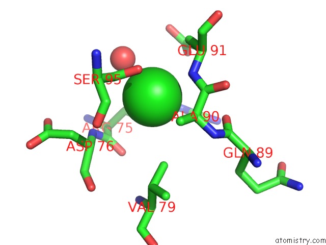
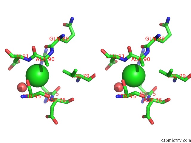
Chlorine binding site 1 out of 3 in 2g9w
Go back to
Chlorine binding site 1 out
of 3 in the Crystal Structure of RV1846C, A Putative Transcriptional Regulatory Protein of Mycobacterium Tuberculosis
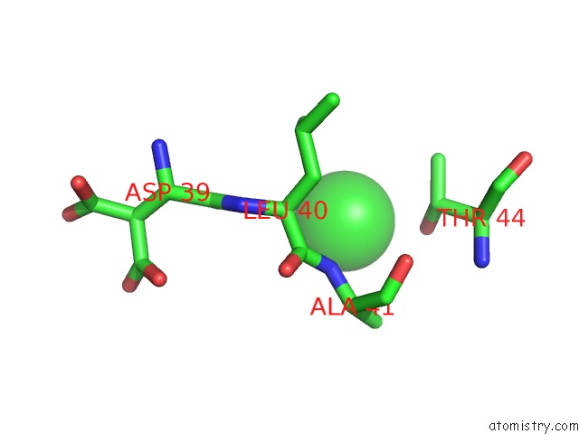
Mono view
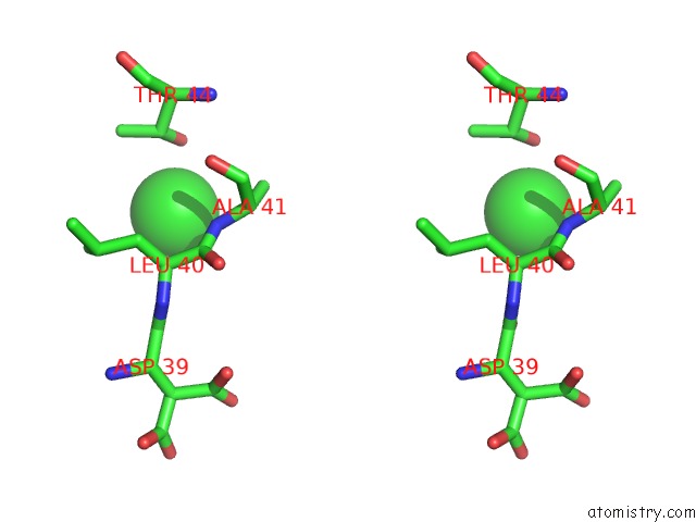
Stereo pair view
Mono view
Stereo pair view
A full contact list of Chlorine with other atoms in the Cl binding
site number 1 of Crystal Structure of RV1846C, A Putative Transcriptional Regulatory Protein of Mycobacterium Tuberculosis within 5.0Å range:
|
Chlorine binding site 2 out of 3 in 2g9w
Go back to
Chlorine binding site 2 out
of 3 in the Crystal Structure of RV1846C, A Putative Transcriptional Regulatory Protein of Mycobacterium Tuberculosis
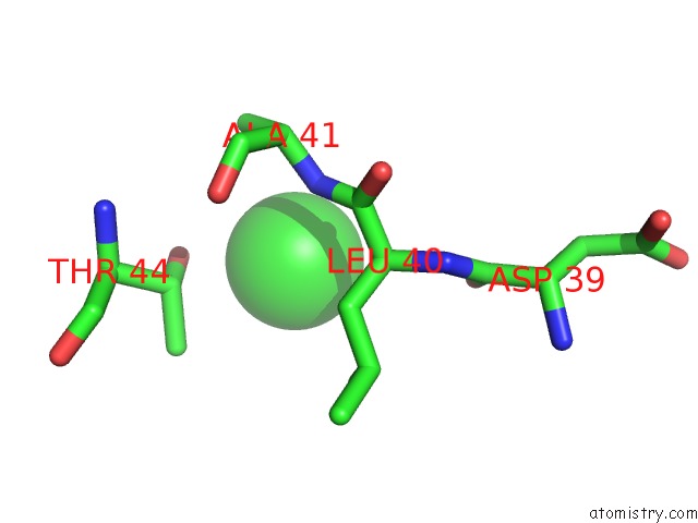
Mono view
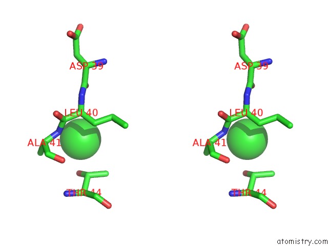
Stereo pair view
Mono view
Stereo pair view
A full contact list of Chlorine with other atoms in the Cl binding
site number 2 of Crystal Structure of RV1846C, A Putative Transcriptional Regulatory Protein of Mycobacterium Tuberculosis within 5.0Å range:
|
Chlorine binding site 3 out of 3 in 2g9w
Go back to
Chlorine binding site 3 out
of 3 in the Crystal Structure of RV1846C, A Putative Transcriptional Regulatory Protein of Mycobacterium Tuberculosis
Mono view
Stereo pair view
Mono view
Stereo pair view
A full contact list of Chlorine with other atoms in the Cl binding
site number 3 of Crystal Structure of RV1846C, A Putative Transcriptional Regulatory Protein of Mycobacterium Tuberculosis within 5.0Å range:
|
Reference:
C.Sala,
A.Haouz,
F.A.Saul,
I.Miras,
I.Rosenkrands,
P.M.Alzari,
S.T.Cole.
Genome-Wide Regulon and Crystal Structure of Blai (RV1846C) From Mycobacterium Tuberculosis Mol.Microbiol. V. 71 1102 2009.
ISSN: ISSN 0950-382X
PubMed: 19154333
DOI: 10.1111/J.1365-2958.2008.06583.X
Page generated: Sat Jul 20 07:24:22 2024
ISSN: ISSN 0950-382X
PubMed: 19154333
DOI: 10.1111/J.1365-2958.2008.06583.X
Last articles
Zn in 9J0NZn in 9J0O
Zn in 9J0P
Zn in 9FJX
Zn in 9EKB
Zn in 9C0F
Zn in 9CAH
Zn in 9CH0
Zn in 9CH3
Zn in 9CH1