Chlorine »
PDB 2g8f-2gm1 »
2gcl »
Chlorine in PDB 2gcl: Structure of the POB3 Middle Domain
Protein crystallography data
The structure of Structure of the POB3 Middle Domain, PDB code: 2gcl
was solved by
A.P.Vandemark,
with X-Ray Crystallography technique. A brief refinement statistics is given in the table below:
| Resolution Low / High (Å) | 30.00 / 2.21 |
| Space group | P 21 21 21 |
| Cell size a, b, c (Å), α, β, γ (°) | 57.110, 57.770, 156.580, 90.00, 90.00, 90.00 |
| R / Rfree (%) | 20.7 / 26.4 |
Chlorine Binding Sites:
The binding sites of Chlorine atom in the Structure of the POB3 Middle Domain
(pdb code 2gcl). This binding sites where shown within
5.0 Angstroms radius around Chlorine atom.
In total 4 binding sites of Chlorine where determined in the Structure of the POB3 Middle Domain, PDB code: 2gcl:
Jump to Chlorine binding site number: 1; 2; 3; 4;
In total 4 binding sites of Chlorine where determined in the Structure of the POB3 Middle Domain, PDB code: 2gcl:
Jump to Chlorine binding site number: 1; 2; 3; 4;
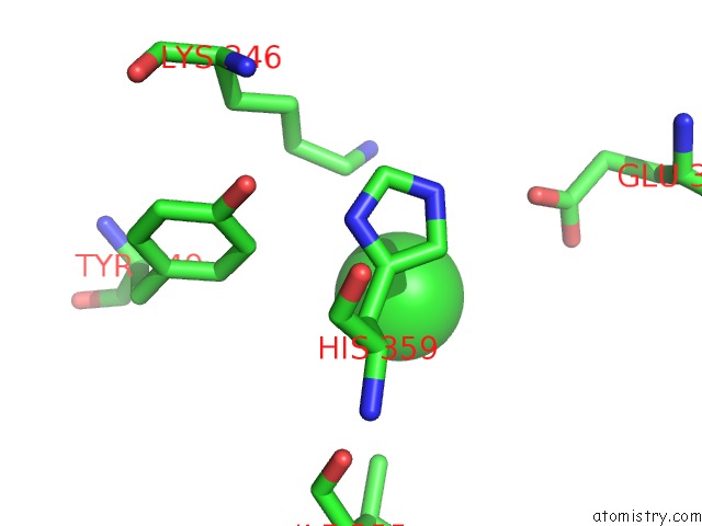
Chlorine binding site 1 out of 4 in 2gcl
Go back to
Chlorine binding site 1 out
of 4 in the Structure of the POB3 Middle Domain
Mono view

Stereo pair view
Mono view
Stereo pair view
A full contact list of Chlorine with other atoms in the Cl binding
site number 1 of Structure of the POB3 Middle Domain within 5.0Å range:
|
Chlorine binding site 2 out of 4 in 2gcl
Go back to
Chlorine binding site 2 out
of 4 in the Structure of the POB3 Middle Domain

Mono view

Stereo pair view
Mono view
Stereo pair view
A full contact list of Chlorine with other atoms in the Cl binding
site number 2 of Structure of the POB3 Middle Domain within 5.0Å range:
|
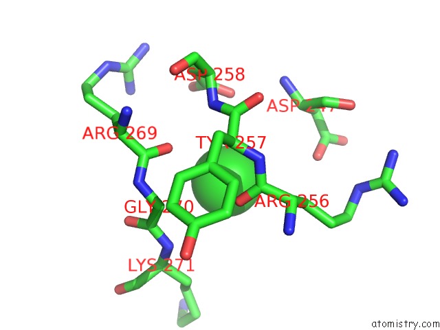
Chlorine binding site 3 out of 4 in 2gcl
Go back to
Chlorine binding site 3 out
of 4 in the Structure of the POB3 Middle Domain

Mono view

Stereo pair view
Mono view
Stereo pair view
A full contact list of Chlorine with other atoms in the Cl binding
site number 3 of Structure of the POB3 Middle Domain within 5.0Å range:
|
Chlorine binding site 4 out of 4 in 2gcl
Go back to
Chlorine binding site 4 out
of 4 in the Structure of the POB3 Middle Domain
Mono view

Stereo pair view
Mono view
Stereo pair view
A full contact list of Chlorine with other atoms in the Cl binding
site number 4 of Structure of the POB3 Middle Domain within 5.0Å range:
|
Reference:
A.P.Vandemark,
M.Blanksma,
E.Ferris,
A.Heroux,
C.P.Hill,
T.Formosa.
The Structure of the Yfact POB3-M Domain, Its Interaction with the Dna Replication Factor Rpa, and A Potential Role in Nucleosome Deposition. Mol.Cell V. 22 363 2006.
ISSN: ISSN 1097-2765
PubMed: 16678108
DOI: 10.1016/J.MOLCEL.2006.03.025
Page generated: Thu Jul 10 22:20:04 2025
ISSN: ISSN 1097-2765
PubMed: 16678108
DOI: 10.1016/J.MOLCEL.2006.03.025
Last articles
F in 4GPBF in 4GS0
F in 4GLU
F in 4GNK
F in 4GOA
F in 4GM8
F in 4GMX
F in 4GLX
F in 4G3B
F in 4GL9