Chlorine »
PDB 2g8f-2gm1 »
2ghw »
Chlorine in PDB 2ghw: Crystal Structure of Sars Spike Protein Receptor Binding Domain in Complex with A Neutralizing Antibody, 80R
Protein crystallography data
The structure of Crystal Structure of Sars Spike Protein Receptor Binding Domain in Complex with A Neutralizing Antibody, 80R, PDB code: 2ghw
was solved by
W.C.Hwang,
Y.Lin,
E.Santelli,
J.Sui,
L.Jaroszewski,
B.Stec,
M.Farzan,
W.A.Marasco,
R.C.Liddington,
with X-Ray Crystallography technique. A brief refinement statistics is given in the table below:
| Resolution Low / High (Å) | 45.55 / 2.30 |
| Space group | P 1 21 1 |
| Cell size a, b, c (Å), α, β, γ (°) | 47.453, 175.902, 67.561, 90.00, 96.55, 90.00 |
| R / Rfree (%) | 24.8 / 29.5 |
Chlorine Binding Sites:
The binding sites of Chlorine atom in the Crystal Structure of Sars Spike Protein Receptor Binding Domain in Complex with A Neutralizing Antibody, 80R
(pdb code 2ghw). This binding sites where shown within
5.0 Angstroms radius around Chlorine atom.
In total 5 binding sites of Chlorine where determined in the Crystal Structure of Sars Spike Protein Receptor Binding Domain in Complex with A Neutralizing Antibody, 80R, PDB code: 2ghw:
Jump to Chlorine binding site number: 1; 2; 3; 4; 5;
In total 5 binding sites of Chlorine where determined in the Crystal Structure of Sars Spike Protein Receptor Binding Domain in Complex with A Neutralizing Antibody, 80R, PDB code: 2ghw:
Jump to Chlorine binding site number: 1; 2; 3; 4; 5;
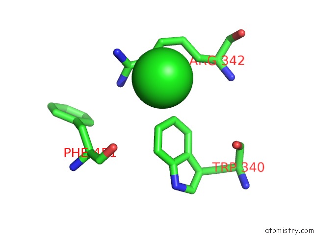
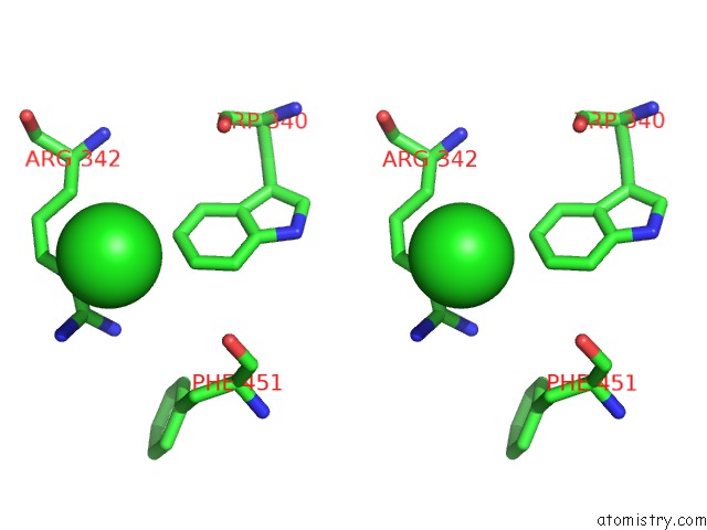
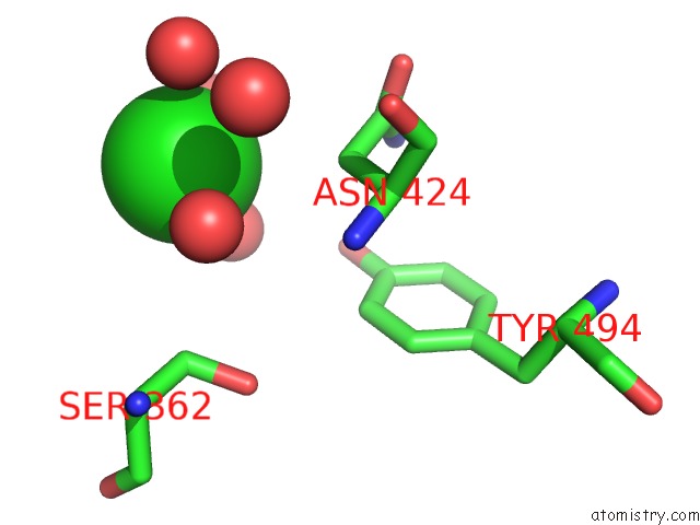
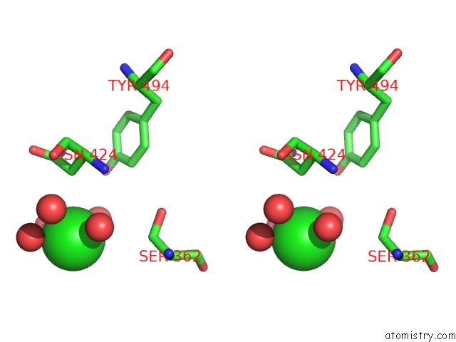
Chlorine binding site 1 out of 5 in 2ghw
Go back to
Chlorine binding site 1 out
of 5 in the Crystal Structure of Sars Spike Protein Receptor Binding Domain in Complex with A Neutralizing Antibody, 80R
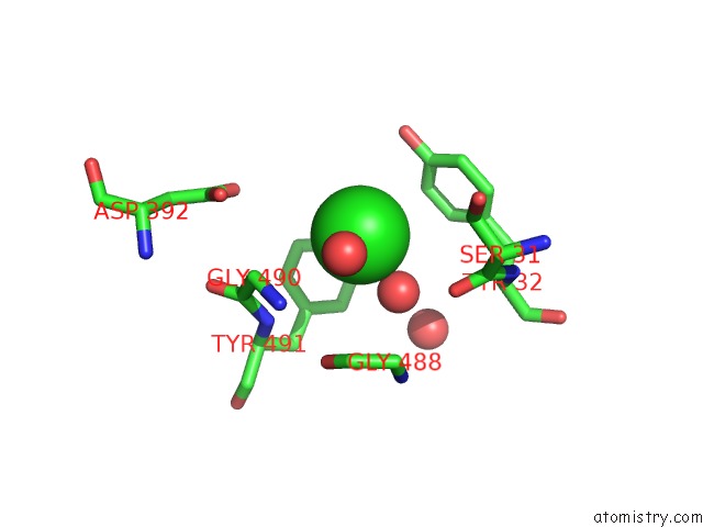
Mono view
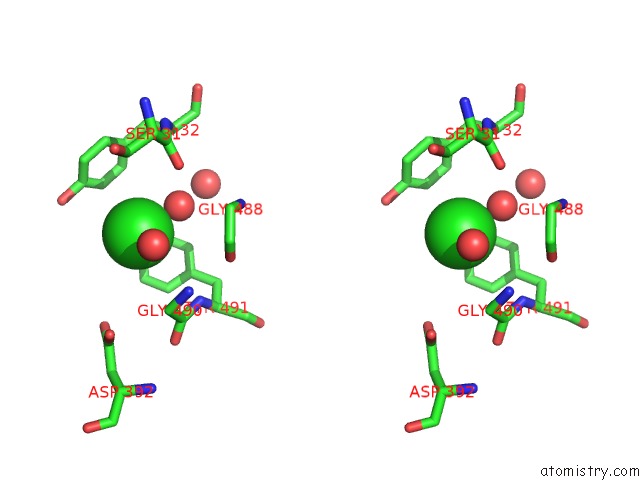
Stereo pair view
Mono view
Stereo pair view
A full contact list of Chlorine with other atoms in the Cl binding
site number 1 of Crystal Structure of Sars Spike Protein Receptor Binding Domain in Complex with A Neutralizing Antibody, 80R within 5.0Å range:
|
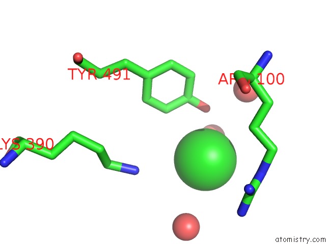
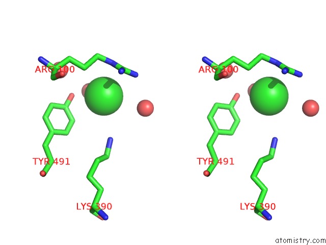
Chlorine binding site 2 out of 5 in 2ghw
Go back to
Chlorine binding site 2 out
of 5 in the Crystal Structure of Sars Spike Protein Receptor Binding Domain in Complex with A Neutralizing Antibody, 80R
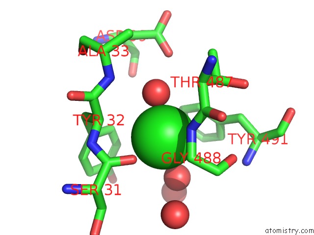
Mono view
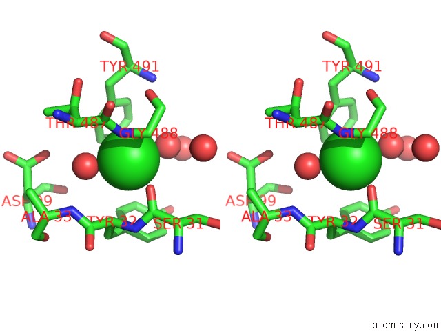
Stereo pair view
Mono view
Stereo pair view
A full contact list of Chlorine with other atoms in the Cl binding
site number 2 of Crystal Structure of Sars Spike Protein Receptor Binding Domain in Complex with A Neutralizing Antibody, 80R within 5.0Å range:
|
Chlorine binding site 3 out of 5 in 2ghw
Go back to
Chlorine binding site 3 out
of 5 in the Crystal Structure of Sars Spike Protein Receptor Binding Domain in Complex with A Neutralizing Antibody, 80R
Mono view
Stereo pair view
Mono view
Stereo pair view
A full contact list of Chlorine with other atoms in the Cl binding
site number 3 of Crystal Structure of Sars Spike Protein Receptor Binding Domain in Complex with A Neutralizing Antibody, 80R within 5.0Å range:
|
Chlorine binding site 4 out of 5 in 2ghw
Go back to
Chlorine binding site 4 out
of 5 in the Crystal Structure of Sars Spike Protein Receptor Binding Domain in Complex with A Neutralizing Antibody, 80R
Mono view
Stereo pair view
Mono view
Stereo pair view
A full contact list of Chlorine with other atoms in the Cl binding
site number 4 of Crystal Structure of Sars Spike Protein Receptor Binding Domain in Complex with A Neutralizing Antibody, 80R within 5.0Å range:
|
Chlorine binding site 5 out of 5 in 2ghw
Go back to
Chlorine binding site 5 out
of 5 in the Crystal Structure of Sars Spike Protein Receptor Binding Domain in Complex with A Neutralizing Antibody, 80R
Mono view
Stereo pair view
Mono view
Stereo pair view
A full contact list of Chlorine with other atoms in the Cl binding
site number 5 of Crystal Structure of Sars Spike Protein Receptor Binding Domain in Complex with A Neutralizing Antibody, 80R within 5.0Å range:
|
Reference:
W.C.Hwang,
Y.Lin,
E.Santelli,
J.Sui,
L.Jaroszewski,
B.Stec,
M.Farzan,
W.A.Marasco,
R.C.Liddington.
Structural Basis of Neutralization By A Human Anti-Severe Acute Respiratory Syndrome Spike Protein Antibody, 80R. J.Biol.Chem. V. 281 34610 2006.
ISSN: ISSN 0021-9258
PubMed: 16954221
DOI: 10.1074/JBC.M603275200
Page generated: Sat Jul 20 07:30:19 2024
ISSN: ISSN 0021-9258
PubMed: 16954221
DOI: 10.1074/JBC.M603275200
Last articles
Zn in 9J0NZn in 9J0O
Zn in 9J0P
Zn in 9FJX
Zn in 9EKB
Zn in 9C0F
Zn in 9CAH
Zn in 9CH0
Zn in 9CH3
Zn in 9CH1