Chlorine »
PDB 2gm9-2h9f »
2gxa »
Chlorine in PDB 2gxa: Crystal Structure of Papillomavirus E1 Hexameric Helicase with Ssdna and Mgadp
Protein crystallography data
The structure of Crystal Structure of Papillomavirus E1 Hexameric Helicase with Ssdna and Mgadp, PDB code: 2gxa
was solved by
E.J.Enemark,
L.Joshua-Tor,
with X-Ray Crystallography technique. A brief refinement statistics is given in the table below:
| Resolution Low / High (Å) | 44.65 / 3.15 |
| Space group | P 1 |
| Cell size a, b, c (Å), α, β, γ (°) | 100.510, 100.881, 125.023, 92.60, 111.46, 106.01 |
| R / Rfree (%) | 23.9 / 29.8 |
Other elements in 2gxa:
The structure of Crystal Structure of Papillomavirus E1 Hexameric Helicase with Ssdna and Mgadp also contains other interesting chemical elements:
| Magnesium | (Mg) | 10 atoms |
Chlorine Binding Sites:
The binding sites of Chlorine atom in the Crystal Structure of Papillomavirus E1 Hexameric Helicase with Ssdna and Mgadp
(pdb code 2gxa). This binding sites where shown within
5.0 Angstroms radius around Chlorine atom.
In total 3 binding sites of Chlorine where determined in the Crystal Structure of Papillomavirus E1 Hexameric Helicase with Ssdna and Mgadp, PDB code: 2gxa:
Jump to Chlorine binding site number: 1; 2; 3;
In total 3 binding sites of Chlorine where determined in the Crystal Structure of Papillomavirus E1 Hexameric Helicase with Ssdna and Mgadp, PDB code: 2gxa:
Jump to Chlorine binding site number: 1; 2; 3;
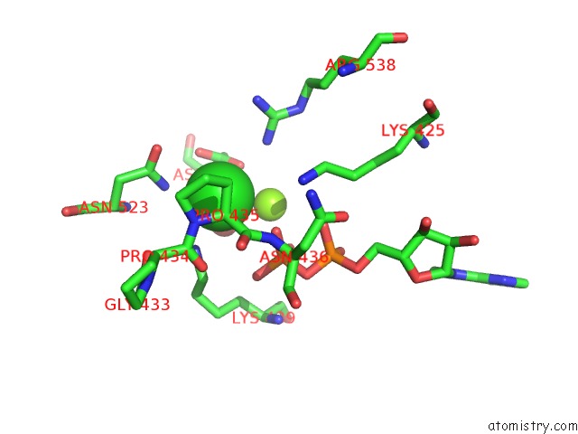
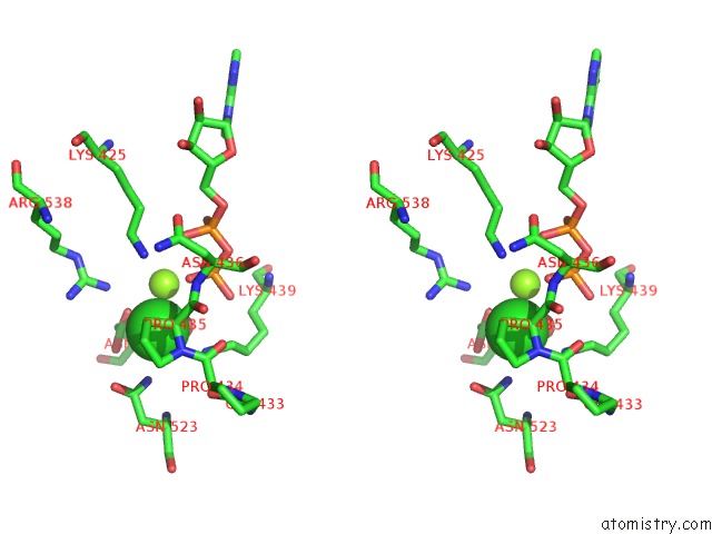
Chlorine binding site 1 out of 3 in 2gxa
Go back to
Chlorine binding site 1 out
of 3 in the Crystal Structure of Papillomavirus E1 Hexameric Helicase with Ssdna and Mgadp
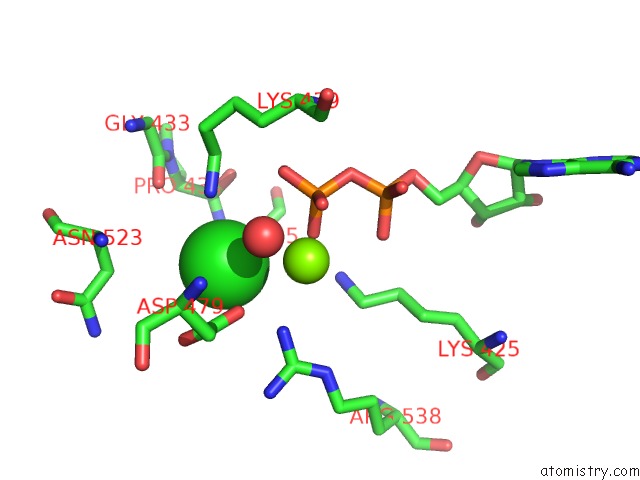
Mono view
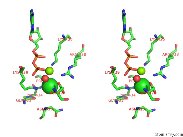
Stereo pair view
Mono view
Stereo pair view
A full contact list of Chlorine with other atoms in the Cl binding
site number 1 of Crystal Structure of Papillomavirus E1 Hexameric Helicase with Ssdna and Mgadp within 5.0Å range:
|
Chlorine binding site 2 out of 3 in 2gxa
Go back to
Chlorine binding site 2 out
of 3 in the Crystal Structure of Papillomavirus E1 Hexameric Helicase with Ssdna and Mgadp
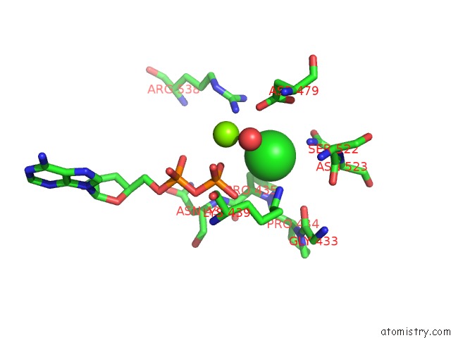
Mono view
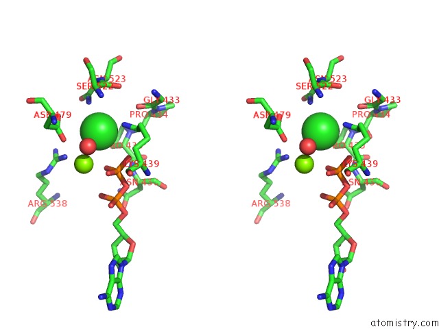
Stereo pair view
Mono view
Stereo pair view
A full contact list of Chlorine with other atoms in the Cl binding
site number 2 of Crystal Structure of Papillomavirus E1 Hexameric Helicase with Ssdna and Mgadp within 5.0Å range:
|
Chlorine binding site 3 out of 3 in 2gxa
Go back to
Chlorine binding site 3 out
of 3 in the Crystal Structure of Papillomavirus E1 Hexameric Helicase with Ssdna and Mgadp
Mono view
Stereo pair view
Mono view
Stereo pair view
A full contact list of Chlorine with other atoms in the Cl binding
site number 3 of Crystal Structure of Papillomavirus E1 Hexameric Helicase with Ssdna and Mgadp within 5.0Å range:
|
Reference:
E.J.Enemark,
L.Joshua-Tor.
Mechanism of Dna Translocation in A Replicative Hexameric Helicase. Nature V. 442 270 2006.
ISSN: ISSN 0028-0836
PubMed: 16855583
DOI: 10.1038/NATURE04943
Page generated: Sat Jul 20 07:41:05 2024
ISSN: ISSN 0028-0836
PubMed: 16855583
DOI: 10.1038/NATURE04943
Last articles
Ca in 5NETCa in 5NER
Ca in 5NEM
Ca in 5NE5
Ca in 5NBP
Ca in 5NBN
Ca in 5NBM
Ca in 5NBL
Ca in 5N7G
Ca in 5N7F