Chlorine »
PDB 2gm1-2h9b »
2h6b »
Chlorine in PDB 2h6b: Crystal Structure of Oxidized Cprk in Complex with O- Chlorophenolacetic Acid
Protein crystallography data
The structure of Crystal Structure of Oxidized Cprk in Complex with O- Chlorophenolacetic Acid, PDB code: 2h6b
was solved by
M.G.Joyce,
C.Levy,
D.Leys,
with X-Ray Crystallography technique. A brief refinement statistics is given in the table below:
| Resolution Low / High (Å) | 19.92 / 2.20 |
| Space group | I 2 2 2 |
| Cell size a, b, c (Å), α, β, γ (°) | 104.437, 112.185, 119.495, 90.00, 90.00, 90.00 |
| R / Rfree (%) | 18.3 / 22.9 |
Chlorine Binding Sites:
The binding sites of Chlorine atom in the Crystal Structure of Oxidized Cprk in Complex with O- Chlorophenolacetic Acid
(pdb code 2h6b). This binding sites where shown within
5.0 Angstroms radius around Chlorine atom.
In total 2 binding sites of Chlorine where determined in the Crystal Structure of Oxidized Cprk in Complex with O- Chlorophenolacetic Acid, PDB code: 2h6b:
Jump to Chlorine binding site number: 1; 2;
In total 2 binding sites of Chlorine where determined in the Crystal Structure of Oxidized Cprk in Complex with O- Chlorophenolacetic Acid, PDB code: 2h6b:
Jump to Chlorine binding site number: 1; 2;
Chlorine binding site 1 out of 2 in 2h6b
Go back to
Chlorine binding site 1 out
of 2 in the Crystal Structure of Oxidized Cprk in Complex with O- Chlorophenolacetic Acid
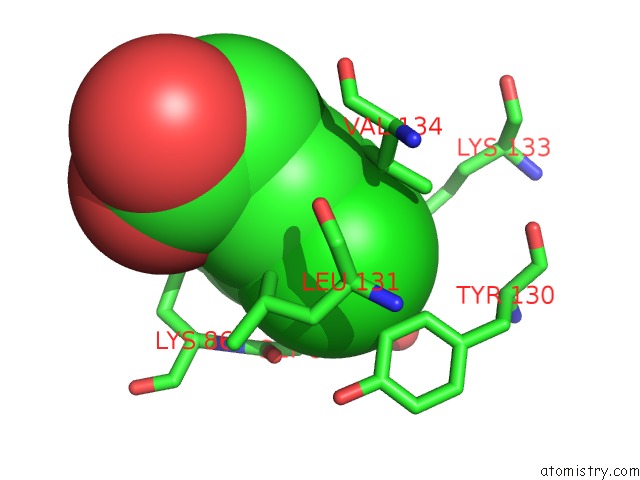
Mono view
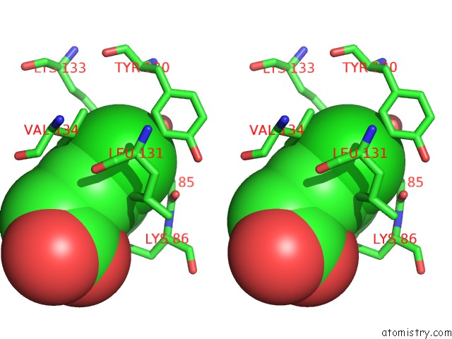
Stereo pair view
Mono view
Stereo pair view
A full contact list of Chlorine with other atoms in the Cl binding
site number 1 of Crystal Structure of Oxidized Cprk in Complex with O- Chlorophenolacetic Acid within 5.0Å range:
|
Chlorine binding site 2 out of 2 in 2h6b
Go back to
Chlorine binding site 2 out
of 2 in the Crystal Structure of Oxidized Cprk in Complex with O- Chlorophenolacetic Acid
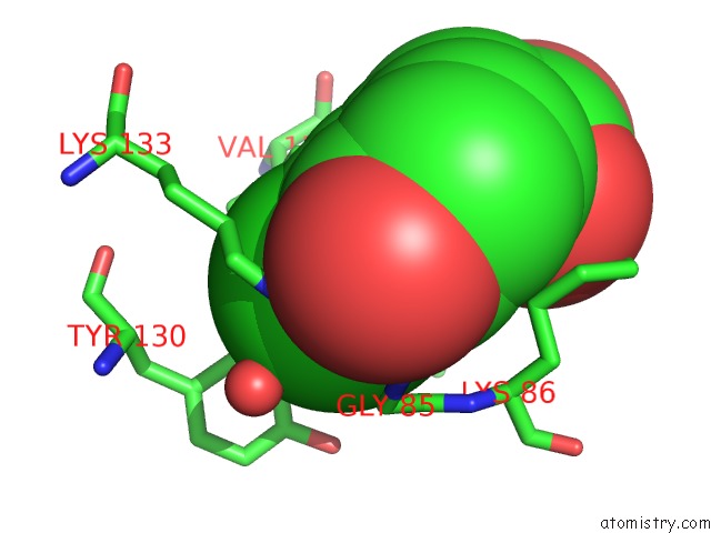
Mono view
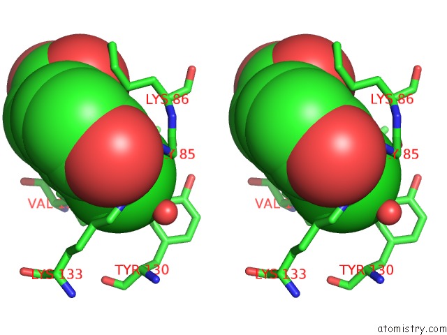
Stereo pair view
Mono view
Stereo pair view
A full contact list of Chlorine with other atoms in the Cl binding
site number 2 of Crystal Structure of Oxidized Cprk in Complex with O- Chlorophenolacetic Acid within 5.0Å range:
|
Reference:
M.G.Joyce,
C.Levy,
S.M.Pop,
B.D.Biehl,
T.I.Doukov,
J.M.Ryter,
H.Mazon,
H.Smidt,
R.H.Van Den Heuvel,
S.W.Ragsdale,
J.Van Der Oost,
D.Leys.
Cprk Crystal Structures Reveal Mechanism For Transcriptional Control of Halorespiration. J.Biol.Chem. V. 281 28318 2006.
ISSN: ISSN 0021-9258
PubMed: 16803881
DOI: 10.1074/JBC.M602654200
Page generated: Sat Jul 20 07:43:25 2024
ISSN: ISSN 0021-9258
PubMed: 16803881
DOI: 10.1074/JBC.M602654200
Last articles
Zn in 9JYWZn in 9IR4
Zn in 9IR3
Zn in 9GMX
Zn in 9GMW
Zn in 9JEJ
Zn in 9ERF
Zn in 9ERE
Zn in 9EGV
Zn in 9EGW