Chlorine »
PDB 2h9j-2hr2 »
2hls »
Chlorine in PDB 2hls: The Crystal Structure of A Protein Disulfide Oxidoreductase From Aeropyrum Pernix K1
Protein crystallography data
The structure of The Crystal Structure of A Protein Disulfide Oxidoreductase From Aeropyrum Pernix K1, PDB code: 2hls
was solved by
K.D'ambrosio,
G.De Simone,
with X-Ray Crystallography technique. A brief refinement statistics is given in the table below:
| Resolution Low / High (Å) | 20.00 / 1.93 |
| Space group | I 2 2 2 |
| Cell size a, b, c (Å), α, β, γ (°) | 90.625, 101.424, 128.927, 90.00, 90.00, 90.00 |
| R / Rfree (%) | 18.7 / 20.9 |
Chlorine Binding Sites:
The binding sites of Chlorine atom in the The Crystal Structure of A Protein Disulfide Oxidoreductase From Aeropyrum Pernix K1
(pdb code 2hls). This binding sites where shown within
5.0 Angstroms radius around Chlorine atom.
In total 2 binding sites of Chlorine where determined in the The Crystal Structure of A Protein Disulfide Oxidoreductase From Aeropyrum Pernix K1, PDB code: 2hls:
Jump to Chlorine binding site number: 1; 2;
In total 2 binding sites of Chlorine where determined in the The Crystal Structure of A Protein Disulfide Oxidoreductase From Aeropyrum Pernix K1, PDB code: 2hls:
Jump to Chlorine binding site number: 1; 2;
Chlorine binding site 1 out of 2 in 2hls
Go back to
Chlorine binding site 1 out
of 2 in the The Crystal Structure of A Protein Disulfide Oxidoreductase From Aeropyrum Pernix K1
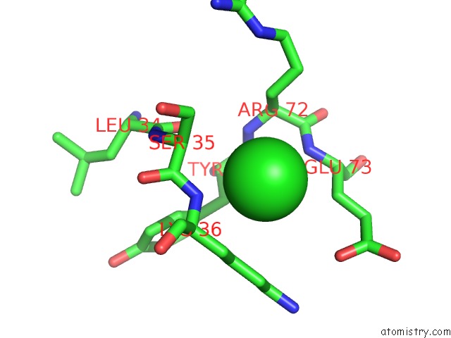
Mono view
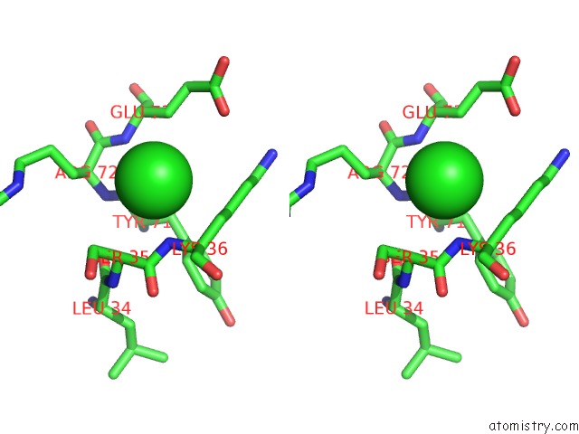
Stereo pair view
Mono view
Stereo pair view
A full contact list of Chlorine with other atoms in the Cl binding
site number 1 of The Crystal Structure of A Protein Disulfide Oxidoreductase From Aeropyrum Pernix K1 within 5.0Å range:
|
Chlorine binding site 2 out of 2 in 2hls
Go back to
Chlorine binding site 2 out
of 2 in the The Crystal Structure of A Protein Disulfide Oxidoreductase From Aeropyrum Pernix K1
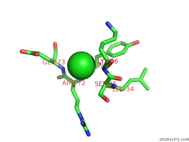
Mono view
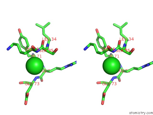
Stereo pair view
Mono view
Stereo pair view
A full contact list of Chlorine with other atoms in the Cl binding
site number 2 of The Crystal Structure of A Protein Disulfide Oxidoreductase From Aeropyrum Pernix K1 within 5.0Å range:
|
Reference:
K.D'ambrosio,
E.Pedone,
E.Langella,
G.De Simone,
M.Rossi,
C.Pedone,
S.Bartolucci.
A Novel Member of the Protein Disulfide Oxidoreductase Family From Aeropyrum Pernix K1: Structure, Function and Electrostatics. J.Mol.Biol. V. 362 743 2006.
ISSN: ISSN 0022-2836
PubMed: 16934838
DOI: 10.1016/J.JMB.2006.07.038
Page generated: Sat Jul 20 07:52:35 2024
ISSN: ISSN 0022-2836
PubMed: 16934838
DOI: 10.1016/J.JMB.2006.07.038
Last articles
Zn in 9J0NZn in 9J0O
Zn in 9J0P
Zn in 9FJX
Zn in 9EKB
Zn in 9C0F
Zn in 9CAH
Zn in 9CH0
Zn in 9CH3
Zn in 9CH1