Chlorine »
PDB 2hrc-2i6f »
2hs1 »
Chlorine in PDB 2hs1: Ultra-High Resolution X-Ray Crystal Structure of Hiv-1 Protease V32I Mutant with TMC114 (Darunavir) Inhibitor
Enzymatic activity of Ultra-High Resolution X-Ray Crystal Structure of Hiv-1 Protease V32I Mutant with TMC114 (Darunavir) Inhibitor
All present enzymatic activity of Ultra-High Resolution X-Ray Crystal Structure of Hiv-1 Protease V32I Mutant with TMC114 (Darunavir) Inhibitor:
3.4.23.16;
3.4.23.16;
Protein crystallography data
The structure of Ultra-High Resolution X-Ray Crystal Structure of Hiv-1 Protease V32I Mutant with TMC114 (Darunavir) Inhibitor, PDB code: 2hs1
was solved by
I.T.Weber,
A.Y.Kovalevsky,
with X-Ray Crystallography technique. A brief refinement statistics is given in the table below:
| Resolution Low / High (Å) | 20.00 / 0.84 |
| Space group | P 21 21 21 |
| Cell size a, b, c (Å), α, β, γ (°) | 28.700, 65.923, 92.534, 90.00, 90.00, 90.00 |
| R / Rfree (%) | 12.4 / 14.9 |
Chlorine Binding Sites:
The binding sites of Chlorine atom in the Ultra-High Resolution X-Ray Crystal Structure of Hiv-1 Protease V32I Mutant with TMC114 (Darunavir) Inhibitor
(pdb code 2hs1). This binding sites where shown within
5.0 Angstroms radius around Chlorine atom.
In total 3 binding sites of Chlorine where determined in the Ultra-High Resolution X-Ray Crystal Structure of Hiv-1 Protease V32I Mutant with TMC114 (Darunavir) Inhibitor, PDB code: 2hs1:
Jump to Chlorine binding site number: 1; 2; 3;
In total 3 binding sites of Chlorine where determined in the Ultra-High Resolution X-Ray Crystal Structure of Hiv-1 Protease V32I Mutant with TMC114 (Darunavir) Inhibitor, PDB code: 2hs1:
Jump to Chlorine binding site number: 1; 2; 3;
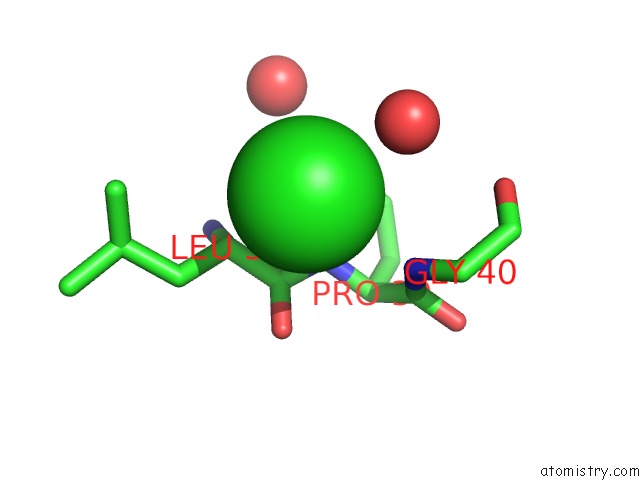
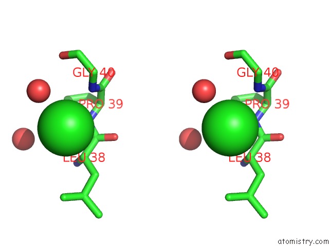
Chlorine binding site 1 out of 3 in 2hs1
Go back to
Chlorine binding site 1 out
of 3 in the Ultra-High Resolution X-Ray Crystal Structure of Hiv-1 Protease V32I Mutant with TMC114 (Darunavir) Inhibitor
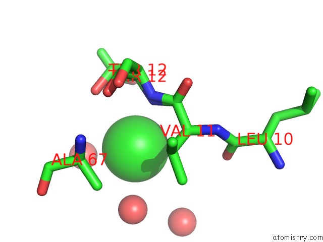
Mono view
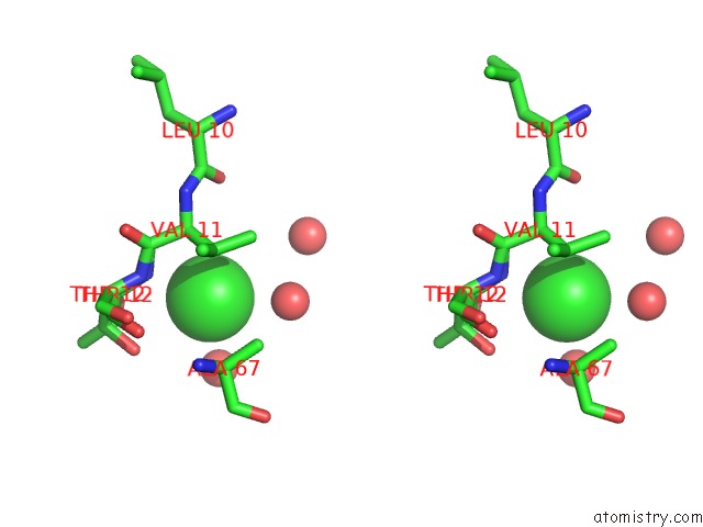
Stereo pair view
Mono view
Stereo pair view
A full contact list of Chlorine with other atoms in the Cl binding
site number 1 of Ultra-High Resolution X-Ray Crystal Structure of Hiv-1 Protease V32I Mutant with TMC114 (Darunavir) Inhibitor within 5.0Å range:
|
Chlorine binding site 2 out of 3 in 2hs1
Go back to
Chlorine binding site 2 out
of 3 in the Ultra-High Resolution X-Ray Crystal Structure of Hiv-1 Protease V32I Mutant with TMC114 (Darunavir) Inhibitor
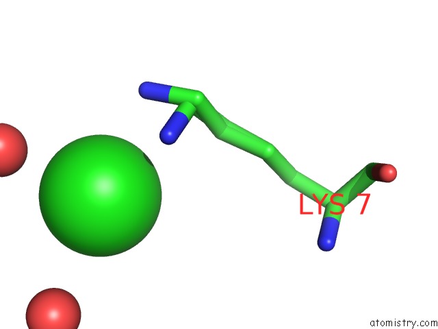
Mono view
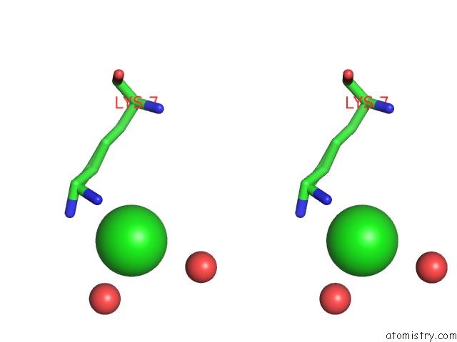
Stereo pair view
Mono view
Stereo pair view
A full contact list of Chlorine with other atoms in the Cl binding
site number 2 of Ultra-High Resolution X-Ray Crystal Structure of Hiv-1 Protease V32I Mutant with TMC114 (Darunavir) Inhibitor within 5.0Å range:
|
Chlorine binding site 3 out of 3 in 2hs1
Go back to
Chlorine binding site 3 out
of 3 in the Ultra-High Resolution X-Ray Crystal Structure of Hiv-1 Protease V32I Mutant with TMC114 (Darunavir) Inhibitor
Mono view
Stereo pair view
Mono view
Stereo pair view
A full contact list of Chlorine with other atoms in the Cl binding
site number 3 of Ultra-High Resolution X-Ray Crystal Structure of Hiv-1 Protease V32I Mutant with TMC114 (Darunavir) Inhibitor within 5.0Å range:
|
Reference:
A.Y.Kovalevsky,
F.Liu,
S.Leshchenko,
A.K.Ghosh,
J.M.Louis,
R.W.Harrison,
I.T.Weber.
Ultra-High Resolution Crystal Structure of Hiv-1 Protease Mutant Reveals Two Binding Sites For Clinical Inhibitor TMC114. J.Mol.Biol. V. 363 161 2006.
ISSN: ISSN 0022-2836
PubMed: 16962136
DOI: 10.1016/J.JMB.2006.08.007
Page generated: Sat Jul 20 07:55:30 2024
ISSN: ISSN 0022-2836
PubMed: 16962136
DOI: 10.1016/J.JMB.2006.08.007
Last articles
Zn in 9MJ5Zn in 9HNW
Zn in 9G0L
Zn in 9FNE
Zn in 9DZN
Zn in 9E0I
Zn in 9D32
Zn in 9DAK
Zn in 8ZXC
Zn in 8ZUF