Chlorine »
PDB 2j6w-2jh6 »
2j91 »
Chlorine in PDB 2j91: Crystal Structure of Human Adenylosuccinate Lyase in Complex with Amp
Enzymatic activity of Crystal Structure of Human Adenylosuccinate Lyase in Complex with Amp
All present enzymatic activity of Crystal Structure of Human Adenylosuccinate Lyase in Complex with Amp:
4.3.2.2;
4.3.2.2;
Protein crystallography data
The structure of Crystal Structure of Human Adenylosuccinate Lyase in Complex with Amp, PDB code: 2j91
was solved by
P.Stenmark,
M.Moche,
C.Arrowsmith,
H.Berglund,
R.Busam,
R.Collins,
A.Edwards,
U.B.Ericsson,
S.Flodin,
A.Flores,
S.Graslund,
M.Hammarstrom,
B.M.Hallberg,
L.Holmberg Schiavone,
M.Hogbom,
I.Johansson,
T.Karlberg,
U.Kosinska,
T.Kotenyova,
A.Magnusdottir,
M.E.Nilsson,
P.Nilsson-Ehle,
T.Nyman,
D.Ogg,
C.Persson,
J.Sagemark,
M.Sundstrom,
J.Uppenberg,
M.Uppsten,
A.G.Thorsell,
S.Van Den Berg,
K.Wallden,
J.Weigelt,
P.Nordlund,
with X-Ray Crystallography technique. A brief refinement statistics is given in the table below:
| Resolution Low / High (Å) | 106.60 / 1.8 |
| Space group | P 21 21 21 |
| Cell size a, b, c (Å), α, β, γ (°) | 85.370, 104.342, 213.396, 90.00, 90.00, 90.00 |
| R / Rfree (%) | 15.7 / 19.9 |
Chlorine Binding Sites:
The binding sites of Chlorine atom in the Crystal Structure of Human Adenylosuccinate Lyase in Complex with Amp
(pdb code 2j91). This binding sites where shown within
5.0 Angstroms radius around Chlorine atom.
In total 4 binding sites of Chlorine where determined in the Crystal Structure of Human Adenylosuccinate Lyase in Complex with Amp, PDB code: 2j91:
Jump to Chlorine binding site number: 1; 2; 3; 4;
In total 4 binding sites of Chlorine where determined in the Crystal Structure of Human Adenylosuccinate Lyase in Complex with Amp, PDB code: 2j91:
Jump to Chlorine binding site number: 1; 2; 3; 4;
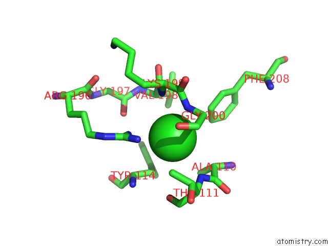
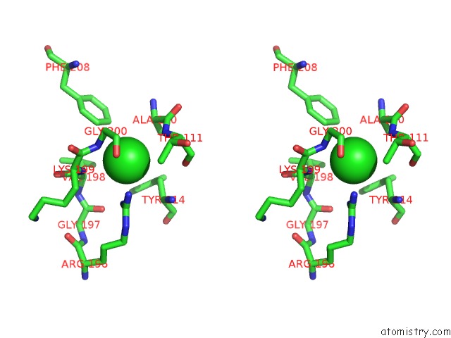
Chlorine binding site 1 out of 4 in 2j91
Go back to
Chlorine binding site 1 out
of 4 in the Crystal Structure of Human Adenylosuccinate Lyase in Complex with Amp
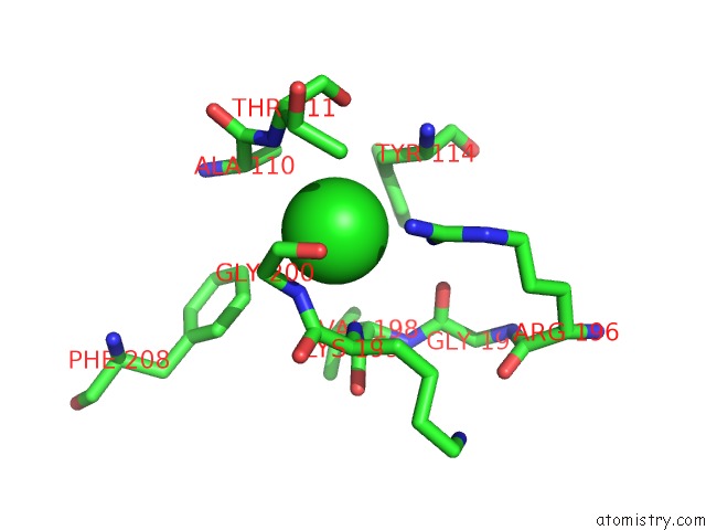
Mono view
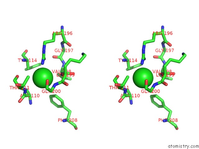
Stereo pair view
Mono view
Stereo pair view
A full contact list of Chlorine with other atoms in the Cl binding
site number 1 of Crystal Structure of Human Adenylosuccinate Lyase in Complex with Amp within 5.0Å range:
|
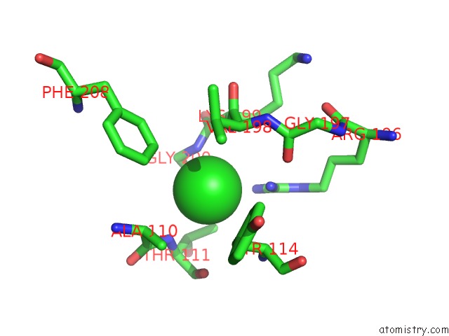
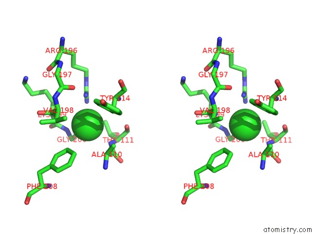
Chlorine binding site 2 out of 4 in 2j91
Go back to
Chlorine binding site 2 out
of 4 in the Crystal Structure of Human Adenylosuccinate Lyase in Complex with Amp
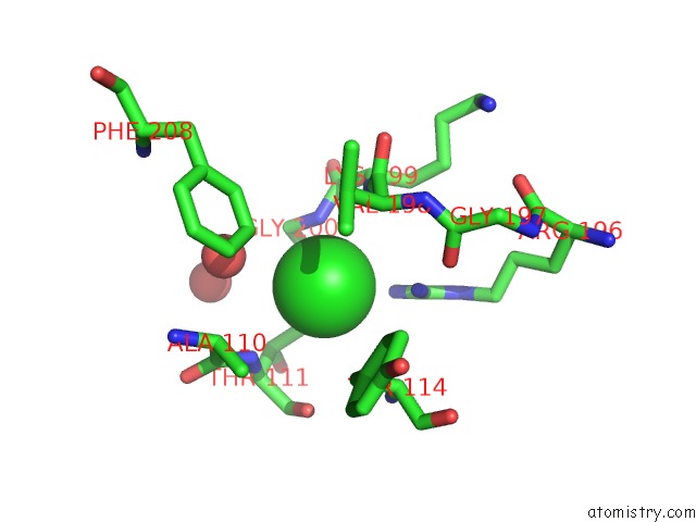
Mono view
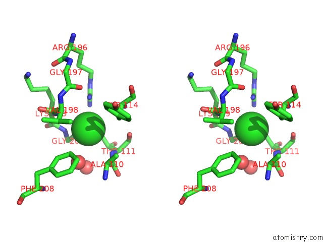
Stereo pair view
Mono view
Stereo pair view
A full contact list of Chlorine with other atoms in the Cl binding
site number 2 of Crystal Structure of Human Adenylosuccinate Lyase in Complex with Amp within 5.0Å range:
|
Chlorine binding site 3 out of 4 in 2j91
Go back to
Chlorine binding site 3 out
of 4 in the Crystal Structure of Human Adenylosuccinate Lyase in Complex with Amp
Mono view
Stereo pair view
Mono view
Stereo pair view
A full contact list of Chlorine with other atoms in the Cl binding
site number 3 of Crystal Structure of Human Adenylosuccinate Lyase in Complex with Amp within 5.0Å range:
|
Chlorine binding site 4 out of 4 in 2j91
Go back to
Chlorine binding site 4 out
of 4 in the Crystal Structure of Human Adenylosuccinate Lyase in Complex with Amp
Mono view
Stereo pair view
Mono view
Stereo pair view
A full contact list of Chlorine with other atoms in the Cl binding
site number 4 of Crystal Structure of Human Adenylosuccinate Lyase in Complex with Amp within 5.0Å range:
|
Reference:
P.Stenmark,
M.Moche,
C.Arrowsmith,
H.Berglund,
R.Busam,
R.Collins,
A.Edwards,
U.B.Ericsson,
S.Flodin,
A.Flores,
S.Graslund,
M.Hammarstrom,
B.M.Hallberg,
L.Holmberg Schiavone,
M.Hogbom,
I.Johansson,
T.Karlberg,
U.Kosinska,
T.Kotenyova,
A.Magnusdottir,
M.E.Nilsson,
P.Nilsson-Ehle,
T.Nyman,
D.Ogg,
C.Persson,
J.Sagemark,
M.Sundstrom,
J.Uppenberg,
M.Uppsten,
A.G.Thorsell,
S.Van Den Berg,
K.Wallden,
J.Weigelt,
P.Nordlund.
Crystal Structure of Human Adenylosuccinate Lyase To Be Published.
Page generated: Sat Jul 20 08:45:07 2024
Last articles
Zn in 9J0NZn in 9J0O
Zn in 9J0P
Zn in 9FJX
Zn in 9EKB
Zn in 9C0F
Zn in 9CAH
Zn in 9CH0
Zn in 9CH3
Zn in 9CH1