Chlorine »
PDB 2kug-2nwh »
2nns »
Chlorine in PDB 2nns: Structure of Inhibitor Binding to Carbonic Anhydrase II
Enzymatic activity of Structure of Inhibitor Binding to Carbonic Anhydrase II
All present enzymatic activity of Structure of Inhibitor Binding to Carbonic Anhydrase II:
4.2.1.1;
4.2.1.1;
Protein crystallography data
The structure of Structure of Inhibitor Binding to Carbonic Anhydrase II, PDB code: 2nns
was solved by
D.W.Christianson,
K.M.Jude,
with X-Ray Crystallography technique. A brief refinement statistics is given in the table below:
| Resolution Low / High (Å) | 50.00 / 1.03 |
| Space group | P 1 21 1 |
| Cell size a, b, c (Å), α, β, γ (°) | 42.264, 41.338, 71.975, 90.00, 104.50, 90.00 |
| R / Rfree (%) | 12.9 / 16.4 |
Other elements in 2nns:
The structure of Structure of Inhibitor Binding to Carbonic Anhydrase II also contains other interesting chemical elements:
| Mercury | (Hg) | 1 atom |
| Zinc | (Zn) | 1 atom |
Chlorine Binding Sites:
The binding sites of Chlorine atom in the Structure of Inhibitor Binding to Carbonic Anhydrase II
(pdb code 2nns). This binding sites where shown within
5.0 Angstroms radius around Chlorine atom.
In total only one binding site of Chlorine was determined in the Structure of Inhibitor Binding to Carbonic Anhydrase II, PDB code: 2nns:
In total only one binding site of Chlorine was determined in the Structure of Inhibitor Binding to Carbonic Anhydrase II, PDB code: 2nns:
Chlorine binding site 1 out of 1 in 2nns
Go back to
Chlorine binding site 1 out
of 1 in the Structure of Inhibitor Binding to Carbonic Anhydrase II
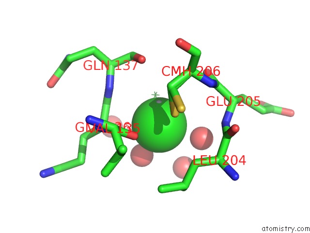
Mono view
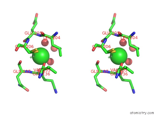
Stereo pair view
Mono view
Stereo pair view
A full contact list of Chlorine with other atoms in the Cl binding
site number 1 of Structure of Inhibitor Binding to Carbonic Anhydrase II within 5.0Å range:
|
Reference:
D.K.Srivastava,
K.M.Jude,
A.L.Banerjee,
M.Haldar,
S.Manokaran,
J.Kooren,
S.Mallik,
D.W.Christianson.
Structural Analysis of Charge Discrimination in the Binding of Inhibitors to Human Carbonic Anhydrases I and II. J.Am.Chem.Soc. V. 129 5528 2007.
ISSN: ISSN 0002-7863
PubMed: 17407288
DOI: 10.1021/JA068359W
Page generated: Sat Jul 20 09:11:01 2024
ISSN: ISSN 0002-7863
PubMed: 17407288
DOI: 10.1021/JA068359W
Last articles
Zn in 9J0NZn in 9J0O
Zn in 9J0P
Zn in 9FJX
Zn in 9EKB
Zn in 9C0F
Zn in 9CAH
Zn in 9CH0
Zn in 9CH3
Zn in 9CH1