Chlorine »
PDB 2nxw-2o9s »
2o4l »
Chlorine in PDB 2o4l: Crystal Structure of Hiv-1 Protease (Q7K, I50V) in Complex with Tipranavir
Protein crystallography data
The structure of Crystal Structure of Hiv-1 Protease (Q7K, I50V) in Complex with Tipranavir, PDB code: 2o4l
was solved by
A.A.Armstrong,
S.Muzammil,
A.Jakalian,
P.R.Bonneau,
V.Schmelmer,
E.Freire,
L.M.Amzel,
with X-Ray Crystallography technique. A brief refinement statistics is given in the table below:
| Resolution Low / High (Å) | 24.35 / 1.33 |
| Space group | P 21 21 2 |
| Cell size a, b, c (Å), α, β, γ (°) | 58.957, 86.411, 46.186, 90.00, 90.00, 90.00 |
| R / Rfree (%) | 19 / 21.6 |
Other elements in 2o4l:
The structure of Crystal Structure of Hiv-1 Protease (Q7K, I50V) in Complex with Tipranavir also contains other interesting chemical elements:
| Fluorine | (F) | 6 atoms |
Chlorine Binding Sites:
The binding sites of Chlorine atom in the Crystal Structure of Hiv-1 Protease (Q7K, I50V) in Complex with Tipranavir
(pdb code 2o4l). This binding sites where shown within
5.0 Angstroms radius around Chlorine atom.
In total 2 binding sites of Chlorine where determined in the Crystal Structure of Hiv-1 Protease (Q7K, I50V) in Complex with Tipranavir, PDB code: 2o4l:
Jump to Chlorine binding site number: 1; 2;
In total 2 binding sites of Chlorine where determined in the Crystal Structure of Hiv-1 Protease (Q7K, I50V) in Complex with Tipranavir, PDB code: 2o4l:
Jump to Chlorine binding site number: 1; 2;
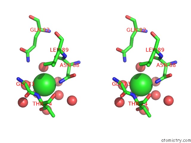
Chlorine binding site 1 out of 2 in 2o4l
Go back to
Chlorine binding site 1 out
of 2 in the Crystal Structure of Hiv-1 Protease (Q7K, I50V) in Complex with Tipranavir

Mono view

Stereo pair view
Mono view
Stereo pair view
A full contact list of Chlorine with other atoms in the Cl binding
site number 1 of Crystal Structure of Hiv-1 Protease (Q7K, I50V) in Complex with Tipranavir within 5.0Å range:
|
Chlorine binding site 2 out of 2 in 2o4l
Go back to
Chlorine binding site 2 out
of 2 in the Crystal Structure of Hiv-1 Protease (Q7K, I50V) in Complex with Tipranavir

Mono view
Stereo pair view
Mono view
Stereo pair view
A full contact list of Chlorine with other atoms in the Cl binding
site number 2 of Crystal Structure of Hiv-1 Protease (Q7K, I50V) in Complex with Tipranavir within 5.0Å range:
|
Reference:
S.Muzammil,
A.A.Armstrong,
L.W.Kang,
A.Jakalian,
P.R.Bonneau,
V.Schmelmer,
L.M.Amzel,
E.Freire.
Unique Thermodynamic Response of Tipranavir to Human Immunodeficiency Virus Type 1 Protease Drug Resistance Mutations. J.Virol. V. 81 5144 2007.
ISSN: ISSN 0022-538X
PubMed: 17360759
DOI: 10.1128/JVI.02706-06
Page generated: Sat Jul 20 09:21:18 2024
ISSN: ISSN 0022-538X
PubMed: 17360759
DOI: 10.1128/JVI.02706-06
Last articles
Zn in 9J0NZn in 9J0O
Zn in 9J0P
Zn in 9FJX
Zn in 9EKB
Zn in 9C0F
Zn in 9CAH
Zn in 9CH0
Zn in 9CH3
Zn in 9CH1