Chlorine »
PDB 2oa1-2ol1 »
2ogi »
Chlorine in PDB 2ogi: Crystal Structure of A Putative Metal Dependent Phosphohydrolase (SAG1661) From Streptococcus Agalactiae Serogroup V at 1.85 A Resolution
Protein crystallography data
The structure of Crystal Structure of A Putative Metal Dependent Phosphohydrolase (SAG1661) From Streptococcus Agalactiae Serogroup V at 1.85 A Resolution, PDB code: 2ogi
was solved by
Joint Center For Structural Genomics (Jcsg),
with X-Ray Crystallography technique. A brief refinement statistics is given in the table below:
| Resolution Low / High (Å) | 40.55 / 1.85 |
| Space group | P 1 21 1 |
| Cell size a, b, c (Å), α, β, γ (°) | 65.841, 52.535, 70.022, 90.00, 114.29, 90.00 |
| R / Rfree (%) | 17.2 / 22.9 |
Other elements in 2ogi:
The structure of Crystal Structure of A Putative Metal Dependent Phosphohydrolase (SAG1661) From Streptococcus Agalactiae Serogroup V at 1.85 A Resolution also contains other interesting chemical elements:
| Iron | (Fe) | 4 atoms |
Chlorine Binding Sites:
The binding sites of Chlorine atom in the Crystal Structure of A Putative Metal Dependent Phosphohydrolase (SAG1661) From Streptococcus Agalactiae Serogroup V at 1.85 A Resolution
(pdb code 2ogi). This binding sites where shown within
5.0 Angstroms radius around Chlorine atom.
In total 3 binding sites of Chlorine where determined in the Crystal Structure of A Putative Metal Dependent Phosphohydrolase (SAG1661) From Streptococcus Agalactiae Serogroup V at 1.85 A Resolution, PDB code: 2ogi:
Jump to Chlorine binding site number: 1; 2; 3;
In total 3 binding sites of Chlorine where determined in the Crystal Structure of A Putative Metal Dependent Phosphohydrolase (SAG1661) From Streptococcus Agalactiae Serogroup V at 1.85 A Resolution, PDB code: 2ogi:
Jump to Chlorine binding site number: 1; 2; 3;
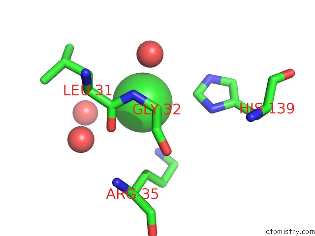
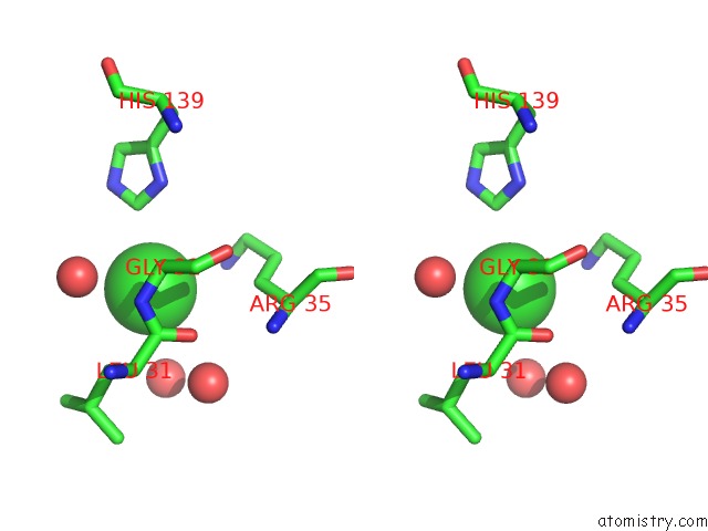
Chlorine binding site 1 out of 3 in 2ogi
Go back to
Chlorine binding site 1 out
of 3 in the Crystal Structure of A Putative Metal Dependent Phosphohydrolase (SAG1661) From Streptococcus Agalactiae Serogroup V at 1.85 A Resolution
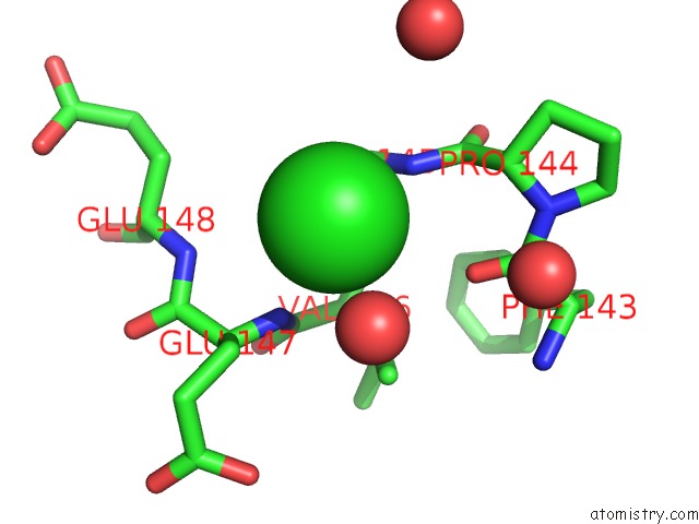
Mono view
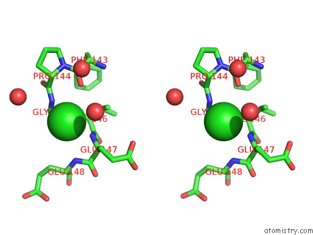
Stereo pair view
Mono view
Stereo pair view
A full contact list of Chlorine with other atoms in the Cl binding
site number 1 of Crystal Structure of A Putative Metal Dependent Phosphohydrolase (SAG1661) From Streptococcus Agalactiae Serogroup V at 1.85 A Resolution within 5.0Å range:
|
Chlorine binding site 2 out of 3 in 2ogi
Go back to
Chlorine binding site 2 out
of 3 in the Crystal Structure of A Putative Metal Dependent Phosphohydrolase (SAG1661) From Streptococcus Agalactiae Serogroup V at 1.85 A Resolution
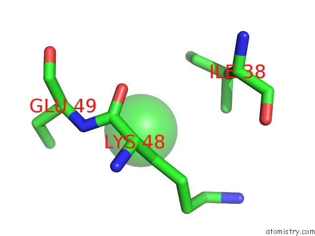
Mono view
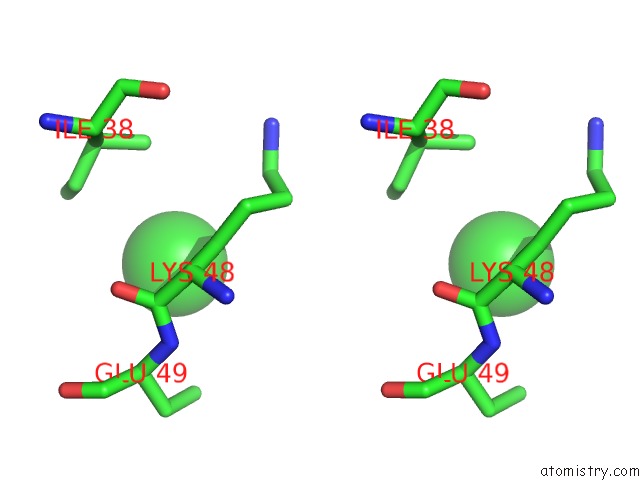
Stereo pair view
Mono view
Stereo pair view
A full contact list of Chlorine with other atoms in the Cl binding
site number 2 of Crystal Structure of A Putative Metal Dependent Phosphohydrolase (SAG1661) From Streptococcus Agalactiae Serogroup V at 1.85 A Resolution within 5.0Å range:
|
Chlorine binding site 3 out of 3 in 2ogi
Go back to
Chlorine binding site 3 out
of 3 in the Crystal Structure of A Putative Metal Dependent Phosphohydrolase (SAG1661) From Streptococcus Agalactiae Serogroup V at 1.85 A Resolution
Mono view
Stereo pair view
Mono view
Stereo pair view
A full contact list of Chlorine with other atoms in the Cl binding
site number 3 of Crystal Structure of A Putative Metal Dependent Phosphohydrolase (SAG1661) From Streptococcus Agalactiae Serogroup V at 1.85 A Resolution within 5.0Å range:
|
Reference:
Joint Center For Structural Genomics (Jcsg),
Joint Center For Structural Genomics (Jcsg).
N/A N/A.
Page generated: Thu Jul 10 23:30:05 2025
Last articles
F in 7NTHF in 7NTI
F in 7NPC
F in 7NRG
F in 7NR5
F in 7NQS
F in 7NOS
F in 7NP5
F in 7NDV
F in 7NP6