Chlorine »
PDB 2ol4-2oud »
2oqx »
Chlorine in PDB 2oqx: Crystal Structure of the Apo Form of E. Coli Tryptophanase at 1.9 A Resolution
Enzymatic activity of Crystal Structure of the Apo Form of E. Coli Tryptophanase at 1.9 A Resolution
All present enzymatic activity of Crystal Structure of the Apo Form of E. Coli Tryptophanase at 1.9 A Resolution:
4.1.99.1;
4.1.99.1;
Protein crystallography data
The structure of Crystal Structure of the Apo Form of E. Coli Tryptophanase at 1.9 A Resolution, PDB code: 2oqx
was solved by
Y.Goldgur,
A.Kogan,
G.Gdalevsky,
A.Parola,
R.Cohen-Luria,
O.Almog,
with X-Ray Crystallography technique. A brief refinement statistics is given in the table below:
| Resolution Low / High (Å) | 14.82 / 1.90 |
| Space group | F 2 2 2 |
| Cell size a, b, c (Å), α, β, γ (°) | 118.422, 120.116, 171.209, 90.00, 90.00, 90.00 |
| R / Rfree (%) | 20.3 / 23.2 |
Other elements in 2oqx:
The structure of Crystal Structure of the Apo Form of E. Coli Tryptophanase at 1.9 A Resolution also contains other interesting chemical elements:
| Magnesium | (Mg) | 1 atom |
Chlorine Binding Sites:
The binding sites of Chlorine atom in the Crystal Structure of the Apo Form of E. Coli Tryptophanase at 1.9 A Resolution
(pdb code 2oqx). This binding sites where shown within
5.0 Angstroms radius around Chlorine atom.
In total only one binding site of Chlorine was determined in the Crystal Structure of the Apo Form of E. Coli Tryptophanase at 1.9 A Resolution, PDB code: 2oqx:
In total only one binding site of Chlorine was determined in the Crystal Structure of the Apo Form of E. Coli Tryptophanase at 1.9 A Resolution, PDB code: 2oqx:
Chlorine binding site 1 out of 1 in 2oqx
Go back to
Chlorine binding site 1 out
of 1 in the Crystal Structure of the Apo Form of E. Coli Tryptophanase at 1.9 A Resolution
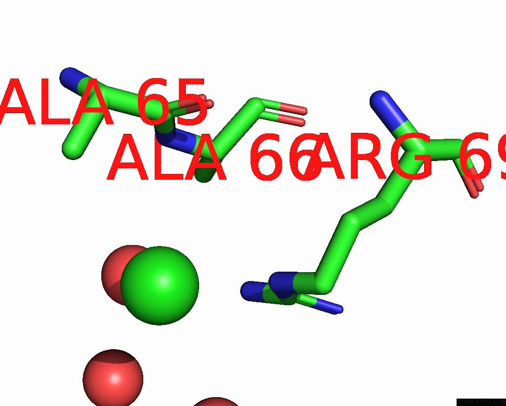
Mono view
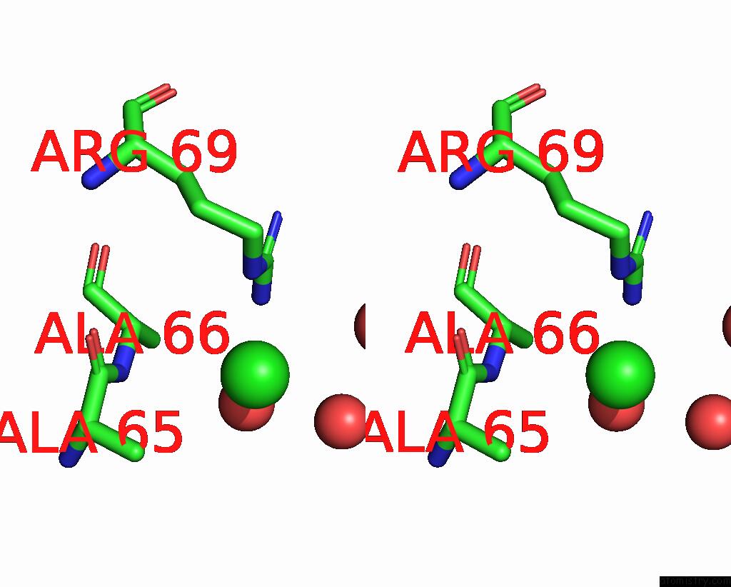
Stereo pair view
Mono view
Stereo pair view
A full contact list of Chlorine with other atoms in the Cl binding
site number 1 of Crystal Structure of the Apo Form of E. Coli Tryptophanase at 1.9 A Resolution within 5.0Å range:
|
Reference:
N.Tsesin,
A.Kogan,
G.Y.Gdalevsky,
J.P.Himanen,
R.Cohen-Luria,
A.H.Parola,
Y.Goldgur,
O.Almog.
The Structure of Apo Tryptophanase From Escherichia Coli Reveals A Wide-Open Conformation. Acta Crystallogr.,Sect.D V. 63 969 2007.
ISSN: ISSN 0907-4449
PubMed: 17704565
DOI: 10.1107/S0907444907036396
Page generated: Sat Jul 20 09:42:37 2024
ISSN: ISSN 0907-4449
PubMed: 17704565
DOI: 10.1107/S0907444907036396
Last articles
Ca in 5M5FCa in 5M3C
Ca in 5M2S
Ca in 5M62
Ca in 5M2O
Ca in 5M11
Ca in 5M0Y
Ca in 5M1P
Ca in 5M27
Ca in 5M0S