Chlorine »
PDB 2oui-2p5v »
2ox0 »
Chlorine in PDB 2ox0: Crystal Structure of JMJD2A Complexed with Histone H3 Peptide Dimethylated at LYS9
Protein crystallography data
The structure of Crystal Structure of JMJD2A Complexed with Histone H3 Peptide Dimethylated at LYS9, PDB code: 2ox0
was solved by
E.S.Pilka,
S.S.Ng,
K.L.Kavanagh,
M.A.Mcdonough,
P.Savitsky,
F.Von Delft,
C.H.Arrowsmith,
J.Weigelt,
A.Edwards,
M.Sundstrom,
C.J.Schofield,
U.Oppermann,
Structural Genomics Consortium (Sgc),
with X-Ray Crystallography technique. A brief refinement statistics is given in the table below:
| Resolution Low / High (Å) | 41.49 / 1.95 |
| Space group | P 21 21 2 |
| Cell size a, b, c (Å), α, β, γ (°) | 101.210, 150.004, 57.228, 90.00, 90.00, 90.00 |
| R / Rfree (%) | 16.9 / 21 |
Other elements in 2ox0:
The structure of Crystal Structure of JMJD2A Complexed with Histone H3 Peptide Dimethylated at LYS9 also contains other interesting chemical elements:
| Nickel | (Ni) | 2 atoms |
| Zinc | (Zn) | 2 atoms |
Chlorine Binding Sites:
The binding sites of Chlorine atom in the Crystal Structure of JMJD2A Complexed with Histone H3 Peptide Dimethylated at LYS9
(pdb code 2ox0). This binding sites where shown within
5.0 Angstroms radius around Chlorine atom.
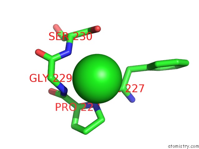
In total only one binding site of Chlorine was determined in the Crystal Structure of JMJD2A Complexed with Histone H3 Peptide Dimethylated at LYS9, PDB code: 2ox0:
In total only one binding site of Chlorine was determined in the Crystal Structure of JMJD2A Complexed with Histone H3 Peptide Dimethylated at LYS9, PDB code: 2ox0:
Chlorine binding site 1 out of 1 in 2ox0
Go back to
Chlorine binding site 1 out
of 1 in the Crystal Structure of JMJD2A Complexed with Histone H3 Peptide Dimethylated at LYS9
Mono view

Stereo pair view
Mono view
Stereo pair view
A full contact list of Chlorine with other atoms in the Cl binding
site number 1 of Crystal Structure of JMJD2A Complexed with Histone H3 Peptide Dimethylated at LYS9 within 5.0Å range:
|
Reference:
S.S.Ng,
K.L.Kavanagh,
M.A.Mcdonough,
D.Butler,
E.S.Pilka,
B.M.Lienard,
J.E.Bray,
P.Savitsky,
O.Gileadi,
F.Von Delft,
N.R.Rose,
J.Offer,
J.C.Scheinost,
T.Borowski,
M.Sundstrom,
C.J.Schofield,
U.Oppermann.
Crystal Structures of Histone Demethylase JMJD2A Reveal Basis For Substrate Specificity. Nature V. 448 87 2007.
ISSN: ISSN 0028-0836
PubMed: 17589501
DOI: 10.1038/NATURE05971
Page generated: Thu Jul 10 23:41:21 2025
ISSN: ISSN 0028-0836
PubMed: 17589501
DOI: 10.1038/NATURE05971
Last articles
Fe in 2YXOFe in 2YRS
Fe in 2YXC
Fe in 2YNM
Fe in 2YVJ
Fe in 2YP1
Fe in 2YU2
Fe in 2YU1
Fe in 2YQB
Fe in 2YOO