Chlorine »
PDB 2p67-2pg4 »
2pc2 »
Chlorine in PDB 2pc2: Lysozyme Cocrystallized with Tris-Dipicolinate Eu Complex
Enzymatic activity of Lysozyme Cocrystallized with Tris-Dipicolinate Eu Complex
All present enzymatic activity of Lysozyme Cocrystallized with Tris-Dipicolinate Eu Complex:
3.2.1.17;
3.2.1.17;
Protein crystallography data
The structure of Lysozyme Cocrystallized with Tris-Dipicolinate Eu Complex, PDB code: 2pc2
was solved by
G.Pompidor,
J.Vicat,
R.Kahn,
with X-Ray Crystallography technique. A brief refinement statistics is given in the table below:
| Resolution Low / High (Å) | 18.98 / 1.54 |
| Space group | C 1 2 1 |
| Cell size a, b, c (Å), α, β, γ (°) | 50.266, 33.779, 69.683, 90.00, 108.22, 90.00 |
| R / Rfree (%) | 13.8 / 16.7 |
Other elements in 2pc2:
The structure of Lysozyme Cocrystallized with Tris-Dipicolinate Eu Complex also contains other interesting chemical elements:
| Europium | (Eu) | 5 atoms |
Chlorine Binding Sites:
The binding sites of Chlorine atom in the Lysozyme Cocrystallized with Tris-Dipicolinate Eu Complex
(pdb code 2pc2). This binding sites where shown within
5.0 Angstroms radius around Chlorine atom.
In total only one binding site of Chlorine was determined in the Lysozyme Cocrystallized with Tris-Dipicolinate Eu Complex, PDB code: 2pc2:
In total only one binding site of Chlorine was determined in the Lysozyme Cocrystallized with Tris-Dipicolinate Eu Complex, PDB code: 2pc2:
Chlorine binding site 1 out of 1 in 2pc2
Go back to
Chlorine binding site 1 out
of 1 in the Lysozyme Cocrystallized with Tris-Dipicolinate Eu Complex
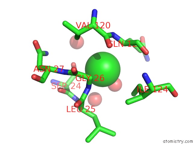
Mono view
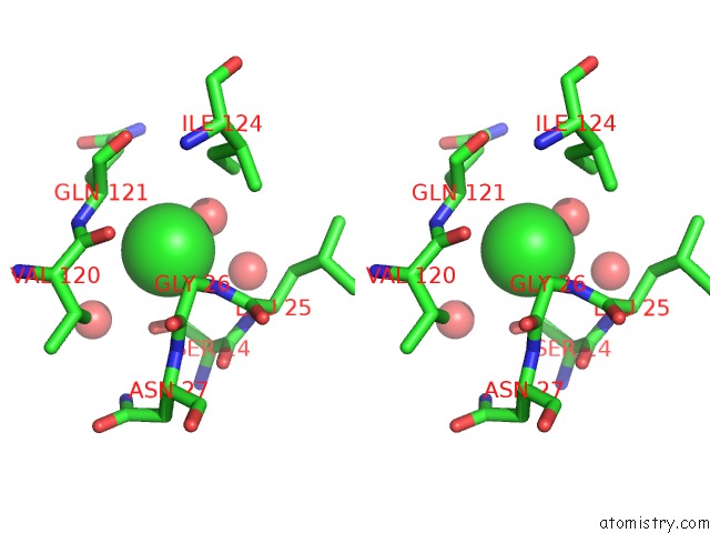
Stereo pair view
Mono view
Stereo pair view
A full contact list of Chlorine with other atoms in the Cl binding
site number 1 of Lysozyme Cocrystallized with Tris-Dipicolinate Eu Complex within 5.0Å range:
|
Reference:
G.Pompidor,
O.Maury,
J.Vicat,
R.Kahn.
A Dipicolinate Lanthanide Complex For Solving Protein Structures Using Anomalous Diffraction Acta Crystallogr.,Sect.D V. 66 762 2010.
ISSN: ISSN 0907-4449
PubMed: 18350532
DOI: 10.1107/S0907444910010954
Page generated: Thu Jul 10 23:48:50 2025
ISSN: ISSN 0907-4449
PubMed: 18350532
DOI: 10.1107/S0907444910010954
Last articles
Fe in 2YXOFe in 2YRS
Fe in 2YXC
Fe in 2YNM
Fe in 2YVJ
Fe in 2YP1
Fe in 2YU2
Fe in 2YU1
Fe in 2YQB
Fe in 2YOO