Chlorine »
PDB 2px4-2q6r »
2pzi »
Chlorine in PDB 2pzi: Crystal Structure of Protein Kinase Pkng From Mycobacterium Tuberculosis in Complex with Tetrahydrobenzothiophene AX20017
Enzymatic activity of Crystal Structure of Protein Kinase Pkng From Mycobacterium Tuberculosis in Complex with Tetrahydrobenzothiophene AX20017
All present enzymatic activity of Crystal Structure of Protein Kinase Pkng From Mycobacterium Tuberculosis in Complex with Tetrahydrobenzothiophene AX20017:
2.7.11.1;
2.7.11.1;
Protein crystallography data
The structure of Crystal Structure of Protein Kinase Pkng From Mycobacterium Tuberculosis in Complex with Tetrahydrobenzothiophene AX20017, PDB code: 2pzi
was solved by
S.Honnappa,
M.O.Steinmetz,
with X-Ray Crystallography technique. A brief refinement statistics is given in the table below:
| Resolution Low / High (Å) | 106.00 / 2.40 |
| Space group | P 65 |
| Cell size a, b, c (Å), α, β, γ (°) | 122.553, 122.553, 243.749, 90.00, 90.00, 120.00 |
| R / Rfree (%) | 18.3 / 23.3 |
Other elements in 2pzi:
The structure of Crystal Structure of Protein Kinase Pkng From Mycobacterium Tuberculosis in Complex with Tetrahydrobenzothiophene AX20017 also contains other interesting chemical elements:
| Cadmium | (Cd) | 2 atoms |
Chlorine Binding Sites:
The binding sites of Chlorine atom in the Crystal Structure of Protein Kinase Pkng From Mycobacterium Tuberculosis in Complex with Tetrahydrobenzothiophene AX20017
(pdb code 2pzi). This binding sites where shown within
5.0 Angstroms radius around Chlorine atom.
In total only one binding site of Chlorine was determined in the Crystal Structure of Protein Kinase Pkng From Mycobacterium Tuberculosis in Complex with Tetrahydrobenzothiophene AX20017, PDB code: 2pzi:
In total only one binding site of Chlorine was determined in the Crystal Structure of Protein Kinase Pkng From Mycobacterium Tuberculosis in Complex with Tetrahydrobenzothiophene AX20017, PDB code: 2pzi:
Chlorine binding site 1 out of 1 in 2pzi
Go back to
Chlorine binding site 1 out
of 1 in the Crystal Structure of Protein Kinase Pkng From Mycobacterium Tuberculosis in Complex with Tetrahydrobenzothiophene AX20017
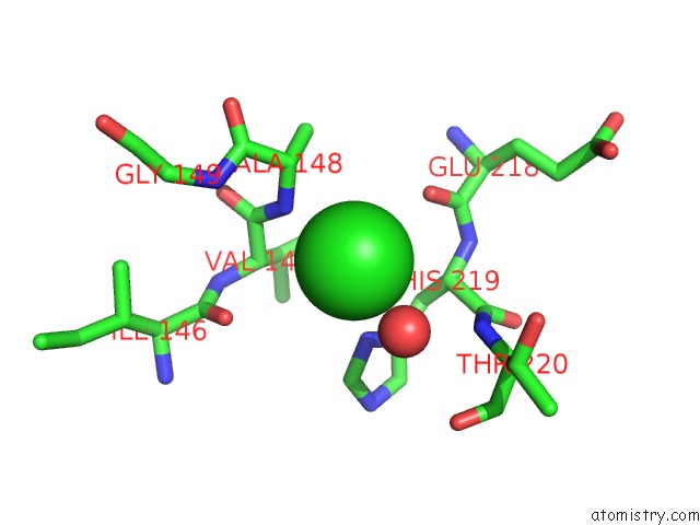
Mono view
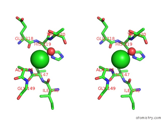
Stereo pair view
Mono view
Stereo pair view
A full contact list of Chlorine with other atoms in the Cl binding
site number 1 of Crystal Structure of Protein Kinase Pkng From Mycobacterium Tuberculosis in Complex with Tetrahydrobenzothiophene AX20017 within 5.0Å range:
|
Reference:
N.Scherr,
S.Honnappa,
G.Kunz,
P.Mueller,
R.Jayachandran,
F.Winkler,
J.Pieters,
M.O.Steinmetz.
Structural Basis For the Specific Inhibition of Protein Kinase G, A Virulence Factor of Mycobacterium Tuberculosis. Proc.Natl.Acad.Sci.Usa V. 104 12151 2007.
ISSN: ISSN 0027-8424
PubMed: 17616581
DOI: 10.1073/PNAS.0702842104
Page generated: Sat Jul 20 10:22:42 2024
ISSN: ISSN 0027-8424
PubMed: 17616581
DOI: 10.1073/PNAS.0702842104
Last articles
Zn in 9J0NZn in 9J0O
Zn in 9J0P
Zn in 9FJX
Zn in 9EKB
Zn in 9C0F
Zn in 9CAH
Zn in 9CH0
Zn in 9CH3
Zn in 9CH1