Chlorine »
PDB 2px4-2q6r »
2q3b »
Chlorine in PDB 2q3b: 1.8 A Resolution Crystal Structure of O-Acetylserine Sulfhydrylase (Oass) Holoenzyme From Mycobacterium Tuberculosis
Enzymatic activity of 1.8 A Resolution Crystal Structure of O-Acetylserine Sulfhydrylase (Oass) Holoenzyme From Mycobacterium Tuberculosis
All present enzymatic activity of 1.8 A Resolution Crystal Structure of O-Acetylserine Sulfhydrylase (Oass) Holoenzyme From Mycobacterium Tuberculosis:
2.5.1.47;
2.5.1.47;
Protein crystallography data
The structure of 1.8 A Resolution Crystal Structure of O-Acetylserine Sulfhydrylase (Oass) Holoenzyme From Mycobacterium Tuberculosis, PDB code: 2q3b
was solved by
G.Schneider,
R.Schnell,
with X-Ray Crystallography technique. A brief refinement statistics is given in the table below:
| Resolution Low / High (Å) | 37.96 / 1.80 |
| Space group | P 41 21 2 |
| Cell size a, b, c (Å), α, β, γ (°) | 70.991, 70.991, 179.624, 90.00, 90.00, 90.00 |
| R / Rfree (%) | 17.4 / 19.2 |
Chlorine Binding Sites:
The binding sites of Chlorine atom in the 1.8 A Resolution Crystal Structure of O-Acetylserine Sulfhydrylase (Oass) Holoenzyme From Mycobacterium Tuberculosis
(pdb code 2q3b). This binding sites where shown within
5.0 Angstroms radius around Chlorine atom.
In total only one binding site of Chlorine was determined in the 1.8 A Resolution Crystal Structure of O-Acetylserine Sulfhydrylase (Oass) Holoenzyme From Mycobacterium Tuberculosis, PDB code: 2q3b:
In total only one binding site of Chlorine was determined in the 1.8 A Resolution Crystal Structure of O-Acetylserine Sulfhydrylase (Oass) Holoenzyme From Mycobacterium Tuberculosis, PDB code: 2q3b:
Chlorine binding site 1 out of 1 in 2q3b
Go back to
Chlorine binding site 1 out
of 1 in the 1.8 A Resolution Crystal Structure of O-Acetylserine Sulfhydrylase (Oass) Holoenzyme From Mycobacterium Tuberculosis
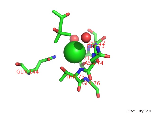
Mono view
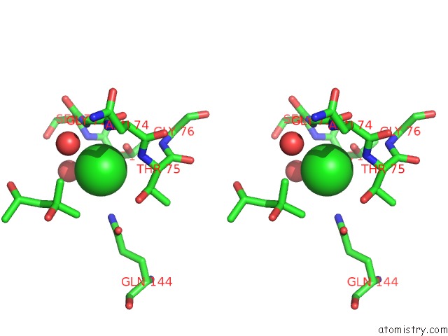
Stereo pair view
Mono view
Stereo pair view
A full contact list of Chlorine with other atoms in the Cl binding
site number 1 of 1.8 A Resolution Crystal Structure of O-Acetylserine Sulfhydrylase (Oass) Holoenzyme From Mycobacterium Tuberculosis within 5.0Å range:
|
Reference:
R.Schnell,
W.Oehlmann,
M.Singh,
G.Schneider.
Structural Insights Into Catalysis and Inhibition of O-Acetylserine Sulfhydrylase From Mycobacterium Tuberculosis: Crystal Structures of the Enzyme {Alpha}-Aminoacrylate Intermediate and An Enzyme-Inhibitor Complex. J.Biol.Chem. V. 282 23473 2007.
ISSN: ISSN 0021-9258
PubMed: 17567578
DOI: 10.1074/JBC.M703518200
Page generated: Sat Jul 20 10:25:18 2024
ISSN: ISSN 0021-9258
PubMed: 17567578
DOI: 10.1074/JBC.M703518200
Last articles
Zn in 9J0NZn in 9J0O
Zn in 9J0P
Zn in 9FJX
Zn in 9EKB
Zn in 9C0F
Zn in 9CAH
Zn in 9CH0
Zn in 9CH3
Zn in 9CH1