Chlorine »
PDB 2qio-2qu5 »
2qrj »
Chlorine in PDB 2qrj: Crystal Structure of Sulfate-Bound Saccharopine Dehydrogenase (L-Lys Forming) From Saccharomyces Cerevisiae
Enzymatic activity of Crystal Structure of Sulfate-Bound Saccharopine Dehydrogenase (L-Lys Forming) From Saccharomyces Cerevisiae
All present enzymatic activity of Crystal Structure of Sulfate-Bound Saccharopine Dehydrogenase (L-Lys Forming) From Saccharomyces Cerevisiae:
1.5.1.7;
1.5.1.7;
Protein crystallography data
The structure of Crystal Structure of Sulfate-Bound Saccharopine Dehydrogenase (L-Lys Forming) From Saccharomyces Cerevisiae, PDB code: 2qrj
was solved by
B.Andi,
H.Xu,
P.F.Cook,
A.H.West,
with X-Ray Crystallography technique. A brief refinement statistics is given in the table below:
| Resolution Low / High (Å) | 30.00 / 1.60 |
| Space group | C 1 2 1 |
| Cell size a, b, c (Å), α, β, γ (°) | 112.628, 55.206, 75.039, 90.00, 116.24, 90.00 |
| R / Rfree (%) | 22.1 / 26 |
Chlorine Binding Sites:
The binding sites of Chlorine atom in the Crystal Structure of Sulfate-Bound Saccharopine Dehydrogenase (L-Lys Forming) From Saccharomyces Cerevisiae
(pdb code 2qrj). This binding sites where shown within
5.0 Angstroms radius around Chlorine atom.
In total only one binding site of Chlorine was determined in the Crystal Structure of Sulfate-Bound Saccharopine Dehydrogenase (L-Lys Forming) From Saccharomyces Cerevisiae, PDB code: 2qrj:
In total only one binding site of Chlorine was determined in the Crystal Structure of Sulfate-Bound Saccharopine Dehydrogenase (L-Lys Forming) From Saccharomyces Cerevisiae, PDB code: 2qrj:
Chlorine binding site 1 out of 1 in 2qrj
Go back to
Chlorine binding site 1 out
of 1 in the Crystal Structure of Sulfate-Bound Saccharopine Dehydrogenase (L-Lys Forming) From Saccharomyces Cerevisiae
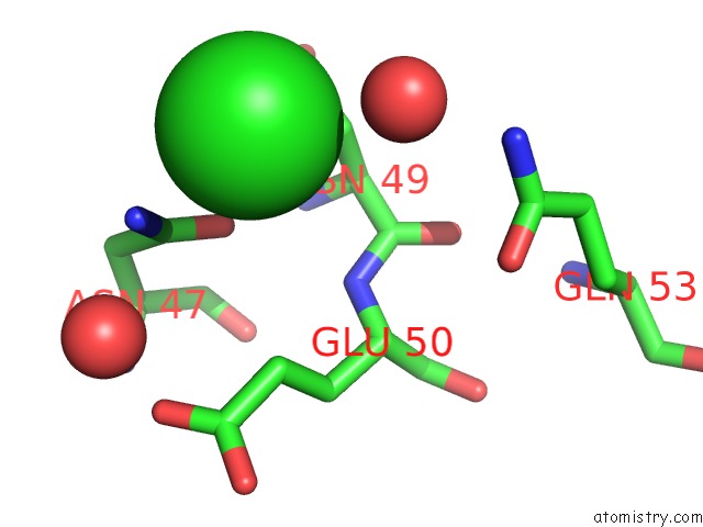
Mono view
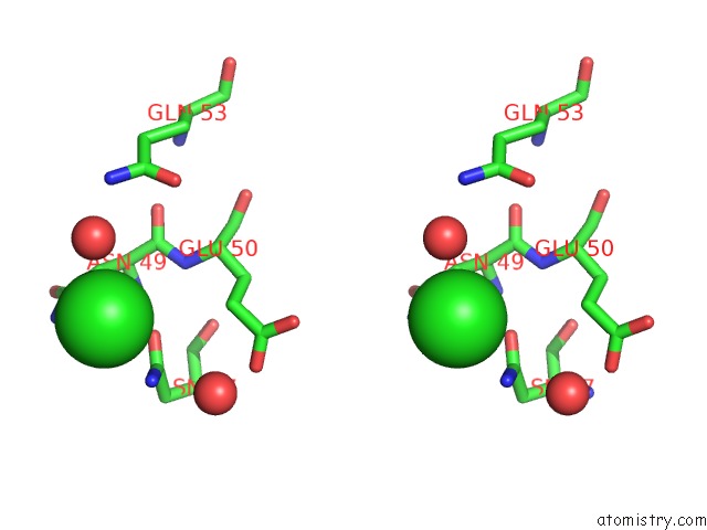
Stereo pair view
Mono view
Stereo pair view
A full contact list of Chlorine with other atoms in the Cl binding
site number 1 of Crystal Structure of Sulfate-Bound Saccharopine Dehydrogenase (L-Lys Forming) From Saccharomyces Cerevisiae within 5.0Å range:
|
Reference:
B.Andi,
H.Xu,
P.F.Cook,
A.H.West.
Crystal Structures of Ligand-Bound Saccharopine Dehydrogenase From Saccharomyces Cerevisiae Biochemistry V. 46 12512 2007.
ISSN: ISSN 0006-2960
PubMed: 17939687
DOI: 10.1021/BI701428M
Page generated: Sat Jul 20 10:52:17 2024
ISSN: ISSN 0006-2960
PubMed: 17939687
DOI: 10.1021/BI701428M
Last articles
Zn in 9J0NZn in 9J0O
Zn in 9J0P
Zn in 9FJX
Zn in 9EKB
Zn in 9C0F
Zn in 9CAH
Zn in 9CH0
Zn in 9CH3
Zn in 9CH1